

Resting-state functional magnetic resonance imaging-based identification of altered brain the fractional amplitude of low frequency fluctuation in adolescent major depressive disorder patients undergoing electroconvulsive therapy
- PMID: 35958635
- PMCID: PMC9357980
- DOI: 10.3389/fpsyt.2022.972968
Resting-state functional magnetic resonance imaging-based identification of altered brain the fractional amplitude of low frequency fluctuation in adolescent major depressive disorder patients undergoing electroconvulsive therapy
Abstract
Purpose: While electroconvulsive therapy (ECT) has been repeatedly been shown to effectively and efficiently treat the major depressive disorder (MDD), the mechanistic basis for such therapeutic efficacy remains to be firmly established. As such, further research exploring the ECT-based treatment of MDD in an adolescent population is warranted.
Methods: This study included 30 treatment-naïve first-episode MDD patients and 30 healthy control (HC) individuals (aged 12-17 years). All participants were scanned using rs-fMRI, and the 30 MDD patients were scanned again after 2 weeks of the ECT treatment period. Intrinsic local activity in each voxel was assessed based on the fractional amplitude of low frequency fluctuation (fALFF) parameter, with all fALFF analyses being completed using the REST application. Correlations between ECT-related changes in fALFF and clinical parameters were additionally examined.
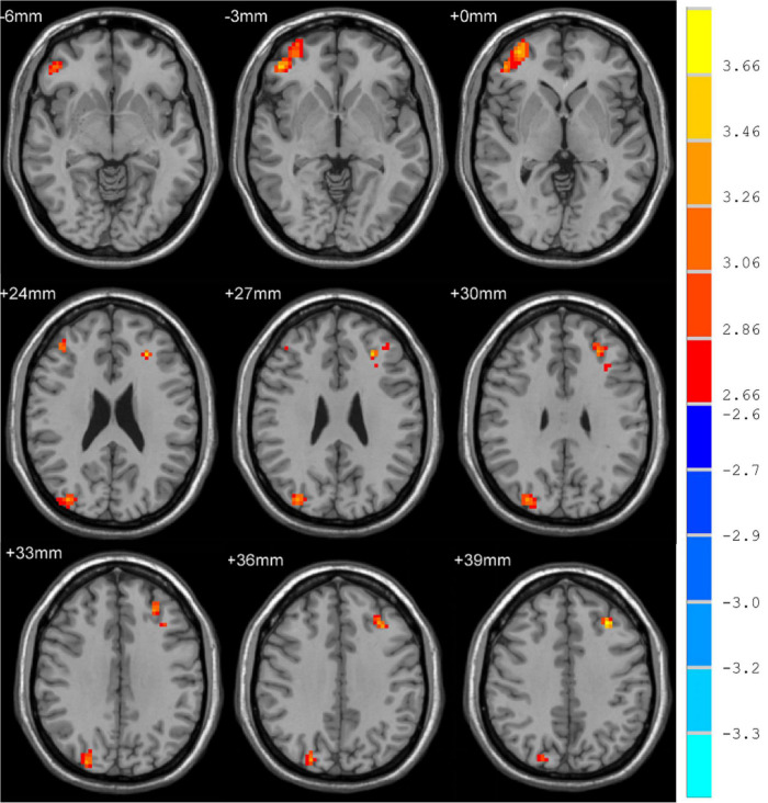
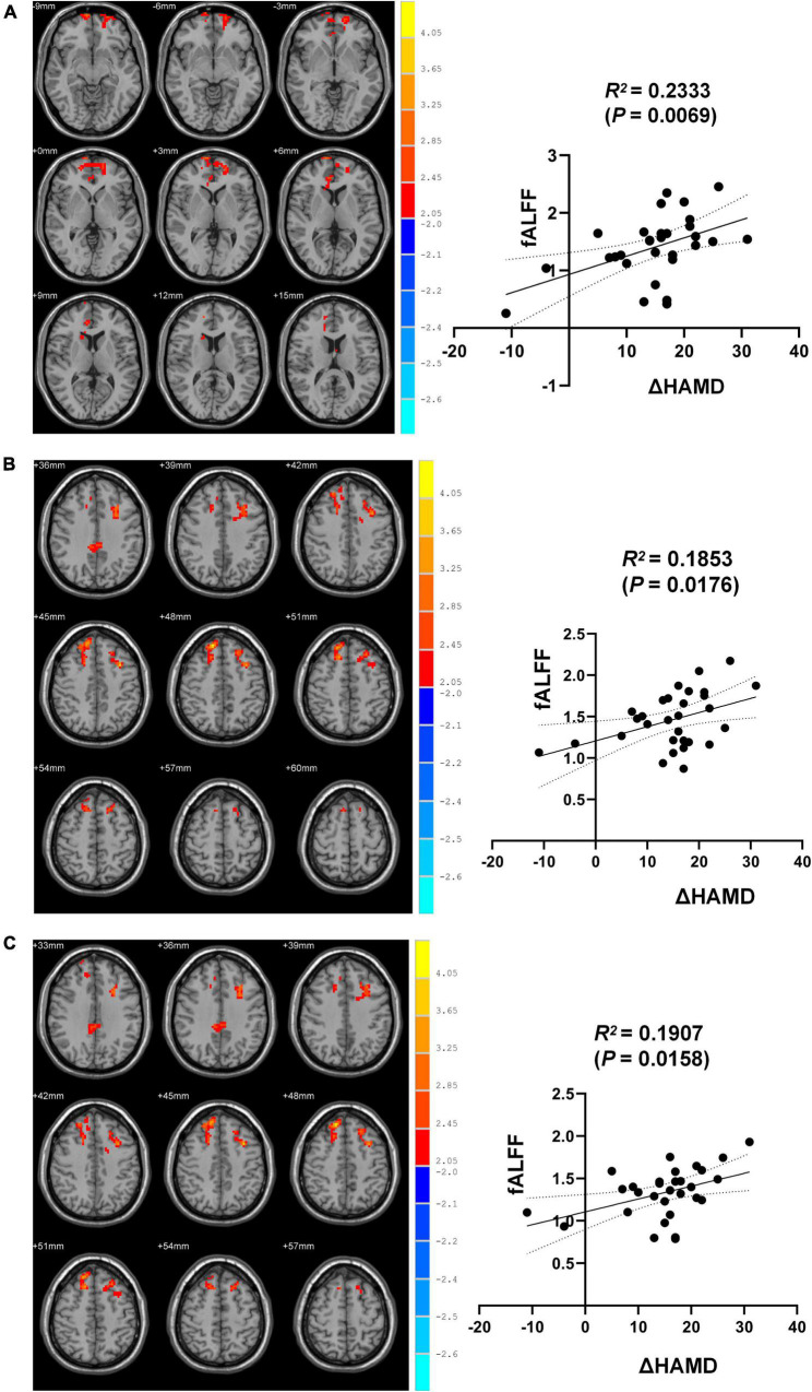
Results: Relative to HCs, MDD patients exhibited increased fALFF values in the right inferior frontal gyrus (ORBinf), inferior occipital gyrus (IOG), and the left middle frontal gyrus (MFG) at baseline. Following ECT, these patients exhibited significant increases in fALFF values in the right medial superior frontal gyrus (SFGmed), dorsolateral superior frontal gyrus (SFGdor), anterior cingulate, and paracingulate gyrus (ACG), median cingulate and paracingulate gyrus (DCG), and left MFG. MDD patient HAMD scores were negatively correlated with fALFF values when analyzing pre-ECT vs. post-HCT ΔHAMD and fALFF values in the right SFGmed, SFGdor, and the left MFG.
Conclusion: These data suggest that ECT induced altered fALFF in some regions of the brain, suggesting that these alterations may serve as a neurobiological indicator of ECT effectiveness in MDD adolescents.
Keywords: adolescent; electroconvulsive therapy; fALFF; major depressive disorder (MDD); resting-state fMRI.
Copyright © 2022 Wang, Tan, Li, Dai, Zhang, Lv and Yu.
Conflict of interest statement
The authors declare that the research was conducted in the absence of any commercial or financial relationships that could be construed as a potential conflict of interest.
Figures
Similar articles
-
Alterations in patients with major depressive disorder before and after electroconvulsive therapy measured by fractional amplitude of low-frequency fluctuations (fALFF).J Affect Disord. 2019 Feb 1;244:92-99. doi: 10.1016/j.jad.2018.10.099. Epub 2018 Oct 9. J Affect Disord. 2019. PMID: 30326347 Free PMC article.
-
Electroconvulsive therapy-induced neuroimaging alterations measured by cerebral blood flow in adolescents with major depressive disorder.J Affect Disord. 2023 Apr 14;327:385-390. doi: 10.1016/j.jad.2023.02.027. Epub 2023 Feb 8. J Affect Disord. 2023. PMID: 36758871 Review.
-
Alteration of Whole Brain ALFF/fALFF and Degree Centrality in Adolescents With Depression and Suicidal Ideation After Electroconvulsive Therapy: A Resting-State fMRI Study.Front Hum Neurosci. 2021 Nov 11;15:762343. doi: 10.3389/fnhum.2021.762343. eCollection 2021. Front Hum Neurosci. 2021. PMID: 34858155 Free PMC article.
-
Altered Fractional Amplitude of Low-Frequency Fluctuation in Major Depressive Disorder and Bipolar Disorder.Front Psychiatry. 2021 Oct 13;12:739210. doi: 10.3389/fpsyt.2021.739210. eCollection 2021. Front Psychiatry. 2021. PMID: 34721109 Free PMC article.
-
Common and distinct patterns of intrinsic brain activity alterations in major depression and bipolar disorder: voxel-based meta-analysis.Transl Psychiatry. 2020 Oct 19;10(1):353. doi: 10.1038/s41398-020-01036-5. Transl Psychiatry. 2020. PMID: 33077728 Free PMC article. Review.
Cited by
-
Hemispheric asymmetries and network dysfunctions in adolescent depression: A neuroimaging study using resting-state functional magnetic resonance imaging.World J Psychiatry. 2025 Feb 19;15(2):102412. doi: 10.5498/wjp.v15.i2.102412. eCollection 2025 Feb 19. World J Psychiatry. 2025. PMID: 39974491 Free PMC article. Clinical Trial.
-
Meta-analysis of resting-state fMRI in cervical spondylosis patients using AES-SDM.Front Neurol. 2024 Sep 24;15:1439939. doi: 10.3389/fneur.2024.1439939. eCollection 2024. Front Neurol. 2024. PMID: 39381074 Free PMC article.
-
Aberrant frontolimbic circuit in female depressed adolescents with and without suicidal attempts: A resting-state functional magnetic resonance imaging study.Front Psychiatry. 2022 Oct 25;13:1007144. doi: 10.3389/fpsyt.2022.1007144. eCollection 2022. Front Psychiatry. 2022. PMID: 36386991 Free PMC article.
-
Longitudinal resting-state network connectivity changes in electroconvulsive therapy patients compared to healthy controls.Brain Stimul. 2024 Jan-Feb;17(1):140-147. doi: 10.1016/j.brs.2023.12.005. Epub 2023 Dec 13. Brain Stimul. 2024. PMID: 38101469 Free PMC article.
References
-
- Huang C, Spuhler K, DeLorenzo C, Parsey R. 604. Association between major depressive disorder and the comt polymorphism as assessed by diffusion MRI. Biol Psychiatry. (2017) 81:S244–5. 10.1016/j.biopsych.2017.02.474 - DOI
LinkOut - more resources
Full Text Sources
Research Materials