

Anatomical Evaluation of Root and Root Canal Morphology of Permanent Mandibular Dentition among the Saudi Arabian Population: A Systematic Review
- PMID: 35958809
- PMCID: PMC9363226
- DOI: 10.1155/2022/2400314
Anatomical Evaluation of Root and Root Canal Morphology of Permanent Mandibular Dentition among the Saudi Arabian Population: A Systematic Review
Abstract
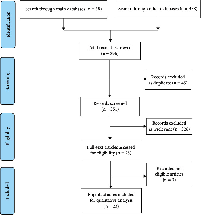
This study aimed to conduct a compendious review of root canal morphology of "permanent mandibular teeth in different regions of Saudi Arabia" to obtain a large sample representing the total population. A detailed search through the databases Web of Science, Scopus, and PubMed was conducted following the PRISMA guidelines. The data were analyzed based on the following inclusion criteria: original full-length original articles that reported the variables of interest "(number of roots, number of canals, Vertucci's classification system and C-shaped canals or mid-mesial canals)" of the mandibular teeth and conducted on Saudi subjects. The retrieved data were presented as frequencies and percentages. The results revealed that 56.6% of mandibular central incisors had one canal and Vertucci type I (56.6%), while 57.4% of the mandibular lateral incisors had one canal, with Vertucci types I and III most frequent. In mandibular canines, 91.8% had one canal and 8.2% had two canals. Most of the mandibular first premolars had one root (86.6%), while almost all mandibular second premolars (91.5%) had one canal, and 96.9% had Vertucci type I configuration. Among the mandibular first molars, three and four canals were prevalent in 58.7% and 40.6%, respectively. The majority of mesial roots had Vertucci type IV (60.6%), and most of distal roots had Vertucci type I (72.2%). Most of the mandibular second molars had three canals (87.3%) and showed Vertucci type IV (39.4%) canals for mesial roots and Vertucci type I (95.6%) for distal roots. The C-shaped canals were seen in 8% of first premolars and 9.8% of second molars. The middle mesial canal was found in 4.2% and 0.4% of first and second molars, respectively. This review could represent "the population of Saudi Arabia as the included samples were combined from different regions of the country." Some variations were noticed within the same group of teeth from different regions. However, the overall results of combined samples were comparable to the other international studies.
Copyright © 2022 Mohammed Mashyakhy et al.
Conflict of interest statement
The authors declare that they have no conflicts of interest.
Figures
References
-
- Mashyakhy M., Chourasia H. R., Jabali A., Almutairi A., Gambarini G. Analysis of fused rooted maxillary first and second molars with merged and C-shaped canal configurations: prevalence, characteristics, and correlations in a Saudi Arabian population. Journal of Endodontia . 2019;45(10):1209–1218. doi: 10.1016/j.joen.2019.06.009. - DOI - PubMed
-
- Mashyakhy M. H., Chourasia H. R., Jabali A. H., et al. C-shaped canal configuration in mandibular premolars and molars: prevalence, correlation, and differences: an in vivo study using cone-beam computed tomography. Nigerian Journal of Clinical Practice . 2020;23(2):232–239. doi: 10.4103/njcp.njcp_335_19. - DOI - PubMed
-
- Margarit R., Andrei O. C. Anatomical variations of mandibular first molar and their implications in endodontic treatment. Romanian Journal of Morphology and Embryology . 2011;52(4):1389–1392. - PubMed
Publication types
MeSH terms
LinkOut - more resources
Full Text Sources
