. 2022 Jul 31:2022:6370766.
doi: 10.1155/2022/6370766. eCollection 2022.

CircAGFG1 Promotes Osteosarcoma Progression and Stemness by Competing with miR-302a-3p to Upregulate the Expression of LATS2

Affiliations

CircAGFG1 Promotes Osteosarcoma Progression and Stemness by Competing with miR-302a-3p to Upregulate the Expression of LATS2

Tongchun Li et al. Evid Based Complement Alternat Med. .

Retraction in

Abstract

This study aimed to investigate the effect of circRNA (circAGFG1) on the proliferation, migration, invasion, and cell stemness of osteosarcoma cells by targeting miR-302a to regulate LATS2. The expression of circAGFG1 in osteosarcoma cells and normal osteoblasts was detected by real-time fluorescent quantitative PCR (RT-qPCR). Cell proliferation, clone formation, and invasion were detected by CCK-8, clone formation, and cell invasion assays. In vivo tumor formation assay was used to detect the effect of circAGFG1 on tumor growth. The expression level of circAGFG1 was upregulated in osteosarcoma cells. The downregulation of circAGFG1 inhibited the proliferation, invasion, and migration of osteosarcoma cells. The overexpression of circAGFG1 enhanced the stemness of osteosarcoma cells. CircAGFG1 was specifically bound to miR-302a to regulate the expression activity of miR-302a. MiR-302a specifically bound to the 3'UTR of LATS2 and inhibited the expression of LATS2. The overexpression of miR-302a reversed the effect of circAGFG1 on the proliferation, invasion, and migration of osteosarcoma cells. CircAGFG1 regulated the expression of LATS2 by miR-302a, thereby regulating the proliferation, migration, and invasion of osteosarcoma cells.


Conflict of interest statement

The authors declare that they have no conflicts of interest.

Figures

Figure 1
Figure 1
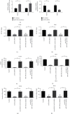
circAGFG1 drives osteosarcoma cell proliferation, migration, invasion, and metastasis. (a) The expression of circAGFG1 in osteosarcoma cell lines was significantly higher than that of FOB and NIH3T3 cells. (b) Detection of transfection efficiency by qRT-PCR. Compared with cells with empty vectors, the use of lentiviral shRNA can significantly reduce the level of circAGFG1. (c) Clone formation assays in U2OS and HOS cells showed that the number of clones was reduced in circAGFG1 knockdown cells compared to control cells. (d) The Matrigel-coated Transwell experiment showed that, when circAGFG1 was knocked down, the invasion potential of U2OS and HOS was significantly reduced. (e) CCK-8 experiment showed that, after knocking down circAGFG1, the proliferation ability of U2OS and HOS was significantly reduced. P < 0.05, ∗∗P < 0.01. NC: negative control. n.s.: not significant.
Figure 2
Figure 2
Knockdown of circAGFG1 inhibits tumor proliferation and metastasis of osteosarcoma. (a) Representative pictures of tumors of HOS cells with different expression levels of circAGFG1 in nude mice. (b) The weight of sh-circAGFG1 xenograft tumor is smaller than that of the control group. (c) The tumor proliferation curve of sh-circAGFG1 xenograft tumor is smaller than that of the control group. (d) Detection of circAGFG1 expression in tumor tissues by qRT-PCR. Compared with cells with empty vectors, the use of lentiviral shRNA can significantly reduce the level of circAGFG1. (e) The tumor metastases of sh-circAGFG1 xenograft tumors were smaller than those of the control group. ∗∗P < 0.01.
Figure 3
Figure 3
Overexpression of circAGFG1 enhances the stemness characteristics of osteosarcoma cells. (a) Detection of transfection efficiency by qRT-PCR. circAGFG1 is highly expressed in the transfected U2OS and HOS cells. (b) In U2OS and HOS cells, overexpression of circAGFG1 upregulated the expression of stemness marker CD133. (c) In U2OS and HOS cells, overexpression of circAGFG1 upregulated the expression of stemness marker SOX2. (d) In U2OS and HOS cells, overexpression of circAGFG1 upregulated the expression of stemness marker CD90. ∗∗P < 0.01.
Figure 4
Figure 4
circAGFG1 as a posttranscriptional regulator. (a) RNA FISH shows that circAGFG1 is mainly located in the cytoplasm of osteosarcoma cells. (b) The predicted potential binding site between circAGFG1 and miR-302a-3p. (c) The dual-luciferase reporter gene verifies the binding of circAGFG1 and miR-302a-3p. Luciferase activity in 293T cells when circAGFG1 WT or Mut vector is cotransfected with miR-302a-3p mimic or negative control. The miR-302a-3p mimic reduced the luciferase activity of the circAGFG1-WT reporter vector but did not reduce the luciferase activity of the empty vector or the mutant reporter vector. (d) The predicted potential binding site between miR-302a-3p and LATS2. (e) The dual-luciferase reporter gene verifies the binding of LATS2 to miR-302a-3p. Luciferase activity in 293T cells when LATS2 WT or Mut vector is cotransfected with miR-302a-3p mimic. The miR-302a-3p mimic reduced the luciferase activity of the LATS2 WT reporter gene vector but did not reduce the luciferase activity of the LATS2 mutant reporter gene vector. (f) Detection of miR-302a-3p expression in tumor tissues by qRT-PCR. Compared with cells with empty vectors, the use of lentivirus shRNA can significantly increase the level of miR-302a-3p. ∗∗P < 0.01. n.s.: not significant.
Figure 5
Figure 5
The correlation of circAGFG1, LATS2, and miR-302a-3p in osteosarcoma cell lines and xenograft tumors. (a) Detection of miR-302a-3p transfection efficiency in U2OS and HOS cells. (b) Detection of LATS2 transfection efficiency in U2OS and HOS cells. (c) U2OS cell proliferation test showed that the cell proliferation ability was significantly reduced in the cells mock-transfected with LATS2-siRNA or miR-302a-3p. The overexpression of circAGFG1 partially reduced the effect of LATS2-siRNA or miR-302a-3p in U2OS cells. (d) The results of cell proliferation assay in HOS cells are similar. (e) U2OS cell clone formation test showed that, in the cells mock-transfected with LATS2-siRNA or miR-302a-3p, the cell clone formation ability was significantly reduced. The overexpression of circAGFG1 partially reduced the effect of LATS2-siRNA or miR-302a-3p in U2OS cells. (f) The results of cell cloning assay in HOS cells are similar. (g) U2OS cell Transwell test showed that in the cells mock-transfected with LATS2-siRNA or miR-302a-3p, the cell migration ability was significantly reduced. The overexpression of circAGFG1 partially reduced the effect of LATS2-siRNA or miR-302a-3p in U2OS cells. (h) The results of cell Transwell assay in HOS cells are similar. ∗∗P < 0.01.
Figure 6
Figure 6
The expression of circAGFG1, LATS2, and miR-302a-3p in osteosarcoma cells and their relationship. (a) The LATS2 mRNA in cells knocked down circAGFG1 was significantly lower than that in control xenograft tumors. (b) The miR-302a-3p in circAGFG1 knockdown cells was significantly higher than that in the control. (c) Compared with control cells, LATS2 is highly expressed in the stably transfected circAGFG1 overexpressing U2OS and HOS cells. (d) Compared with control cells, miR-302a-3p is underexpressed in stably transfected circAGFG1 overexpressing U2OS and HOS cells. (e) In U2OS and HOS cells, overexpression of miR-302a-3p inhibits the expression of LATS2. ∗∗P < 0.01.

Similar articles

Cited by

References

    1. Misaghi A., Goldin A., Awad M., Kulidjian A. A. Osteosarcoma: a comprehensive review. SICOT-J . 2018;4:p. 12. doi: 10.1051/sicotj/2017028. - DOI - PMC - PubMed
    1. Sayles L. C., Breese M. R., Koehne A. L., et al. Genome-informed targeted therapy for osteosarcoma. Cancer Discovery . 2019;9(1):46–63. doi: 10.1158/2159-8290.cd-17-1152. - DOI - PMC - PubMed
    1. Harrison D. J., Geller D. S., Gill J. D., Lewis V. O., Gorlick R. Current and future therapeutic approaches for osteosarcoma. Expert Review of Anticancer Therapy . 2018;18(1):39–50. doi: 10.1080/14737140.2018.1413939. - DOI - PubMed
    1. Zhang Y., Yang J., Zhao N., et al. Progress in the chemotherapeutic treatment of osteosarcoma. Oncology Letters . 2018;16(5):6228–6237. doi: 10.3892/ol.2018.9434. - DOI - PMC - PubMed
    1. Smrke A., Anderson P. M., Gulia A., Gennatas S., Huang P. H., Jones R. L. Future directions in the treatment of osteosarcoma. Cells . 2021;10(1) doi: 10.3390/cells10010172. - DOI - PMC - PubMed

Publication types

LinkOut - more resources