Review
. 2022 Mar 31:3:835503.
doi: 10.3389/falgy.2022.835503. eCollection 2022.

SERPING1 Variants and C1-INH Biological Function: A Close Relationship With C1-INH-HAE

Affiliations
Review

SERPING1 Variants and C1-INH Biological Function: A Close Relationship With C1-INH-HAE

Christian Drouet et al. Front Allergy. .

Abstract

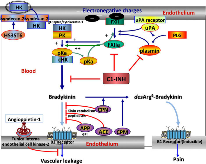
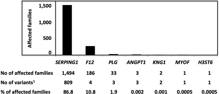
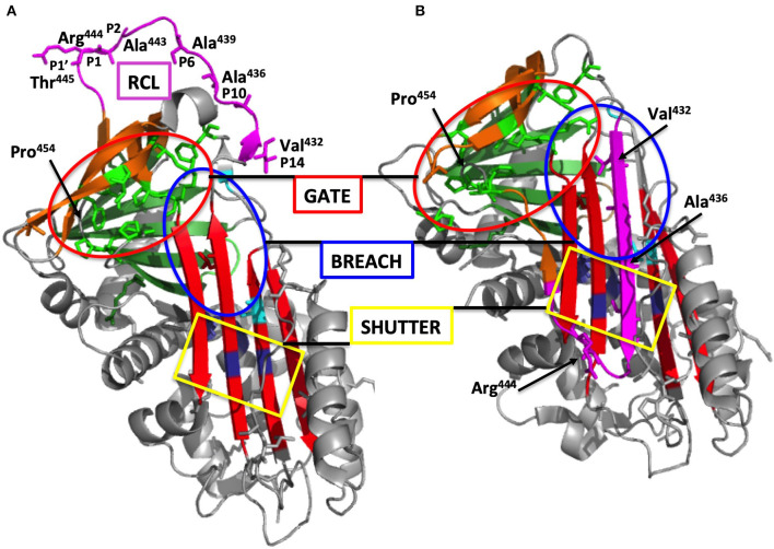
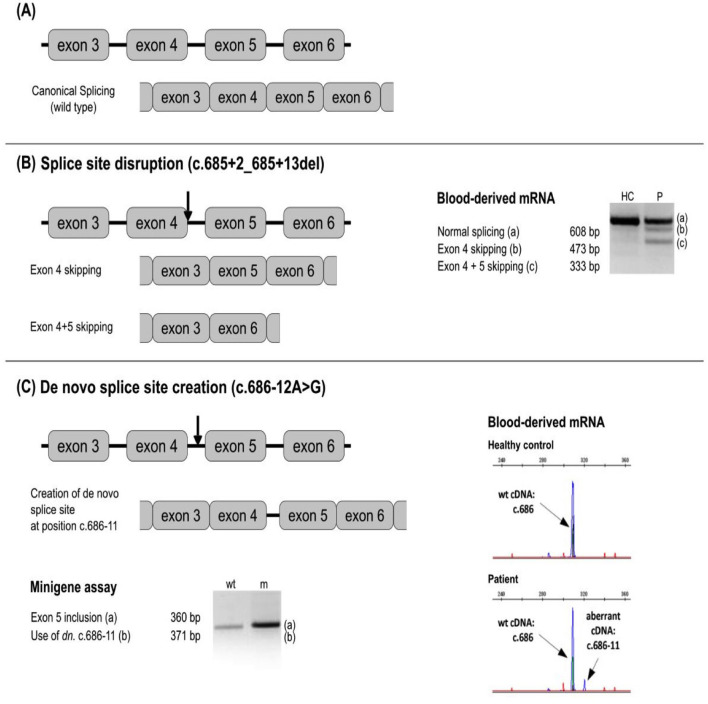
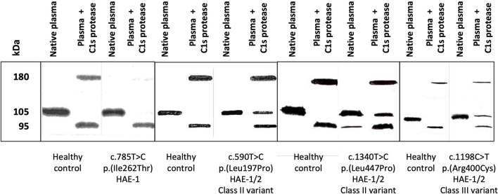
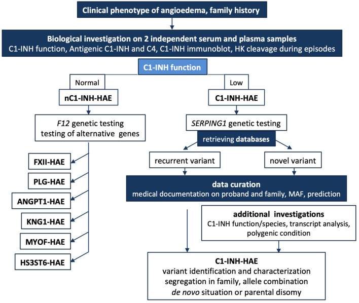
Hereditary angioedema with C1 Inhibitor deficiency (C1-INH-HAE) is caused by a constellation of variants of the SERPING1 gene (n = 809; 1,494 pedigrees), accounting for 86.8% of HAE families, showing a pronounced mutagenic liability of SERPING1 and pertaining to 5.6% de novo variants. C1-INH is the major control serpin of the kallikrein-kinin system (KKS). In addition, C1-INH controls complement C1 and plasminogen activation, both systems contributing to inflammation. Recognizing the failed control of C1s protease or KKS provides the diagnosis of C1-INH-HAE. SERPING1 variants usually behave in an autosomal-dominant character with an incomplete penetrance and a low prevalence. A great majority of variants (809/893; 90.5%) that were introduced into online database have been considered as pathogenic/likely pathogenic. Haploinsufficiency is a common feature in C1-INH-HAE where a dominant-negative variant product impacts the wild-type allele and renders it inactive. Small (36.2%) and large (8.3%) deletions/duplications are common, with exon 4 as the most affected one. Point substitutions with missense variants (32.2%) are of interest for the serpin structure-function relationship. Canonical splice sites can be affected by variants within introns and exons also (14.3%). For noncanonical sequences, exon skipping has been confirmed by splicing analyses of patients' blood-derived RNAs (n = 25). Exonic variants (n = 6) can affect exon splicing. Rare deep-intron variants (n = 6), putatively acting as pseudo-exon activating mutations, have been characterized as pathogenic. Some variants have been characterized as benign/likely benign/of uncertain significance (n = 74). This category includes some homozygous (n = 10) or compound heterozygous variants (n = 11). They are presenting with minor allele frequency (MAF) below 0.00002 (i.e., lower than C1-INH-HAE frequency), and may be quantitatively unable to cause haploinsufficiency. Rare benign variants could contribute as disease modifiers. Gonadal mosaicism in C1-INH-HAE is rare and must be distinguished from a de novo variant. Situations with paternal or maternal disomy have been recorded (n = 3). Genotypes must be interpreted with biological investigation fitting with C1-INH expression and typing. Any SERPING1 variant reminiscent of the dysfunctional phenotype of serpin with multimerization or latency should be identified as serpinopathy.

Keywords: C1 Inhibitor; C1-INH-HAE; SERPING1 gene; angioedema; genetic variation; hereditary–diagnosis; serpin function; serpinopathy.


Conflict of interest statement

AGh was employed by KininX SAS. FP and AGe were employed by CeMIA SA. The remaining authors declare that the research was conducted in the absence of any commercial or financial relationships that could be construed as a potential conflict of interest.

Figures

Figure 1
Figure 1
The central position of C1 Inhibitor (C1-INH) within the kallikrein–kinin system (KKS) and its companions. KKS activation is triggered when FXII becomes activated into FXIIa on binding to an activator onto an electronegative membrane. KKS activation results in HK cHK and bradykinin (BK). The serpin C1-INH controls both activation and activity of KKS, with zymogen to enzyme conversion of FXII and pK. C1-INH also controls the reciprocal activation of FXII and pK by plasmin in an interconnected amplification (5). ANGPT1 is a secreted protein ligand for tunica interna endothelium kinase-2, a receptor expressed in growing vascular endothelial cells. ANGPT1 targets key mechanisms contributing to the maintenance of endothelium function by inhibiting the effects of permeability enhancing agents, including BK, protecting from extensive permeability. H3ST6 is involved in HK docking on the endothelial cell surface, preventing HK binding to gC1q receptor and cytokeratin-1 and engaging in kinin forming. Aminopeptidase P (APP) and Angiotensin-I converting enzyme (ACE) are the membrane peptidases that inactivate BK, whereas carboxypeptidases M and N transform a B2 ligand into a B1 ligand. What this scheme means for understanding hereditary angioedema (HAE). SERPING1 variants display a markedly reduced control function of C1-INH toward KKS and plasmin. F12 and PLG variants are shown to increase FXII and plasminogen (PLG) activation, respectively, and KNG1 variants more susceptible to HK cleavage with BK production. ANGPT1 variants showed reduced capacity to bind its natural receptor, with less control of BK-dependent vascular leakage. Because of incomplete heparan-sulfate modification of syndecan-2 by H3ST6, H3ST6 variants were less able to take up HK via endocytosis into the endothelial cell and more HKs entering into the KKS process. Dark blue arrows indicate activation, green arrows the amplification loop, light blue arrows the ligand–receptor interactions, and red lines indicate an inactivation of BK function. FXII, Factor XII; pK, prekallikrein; pKa, plasma kallikrein; HK, High-molecular-weight kininogen; cHK, cleaved HK; ANGPT1, angiopoietin-1; H3ST6, Heparan-sulfate-glucosamine 3-O-sulfotransferase 6; uPA, urokinase-type plasminogen activator.
Figure 2
Figure 2
The distribution of variants responsible for HAE and characterized as pathogenic/likely pathogenic, with a number of affected families. Pathogenic/likely pathogenic variants have been characterized in agreement with the American College of Medical Genetics (ACMG) criteria or as declared by authors in the observations. 1Variants were characterized as pathogenic/likely pathogenic.
Figure 3
Figure 3
Overall structure of C1-INH. (A) Native C1-INH serpin domain, with positions of strategic residues shown with side chains in sticks. Five regions for the serpin function are presented on the 3D-model of C1Inh (PDB ID 5DU3) presented using Pymol. The model starts at Phe100 and lacks a great part of the N-terminal domain (residues 1-112). Strategic functional regions are indicated with (i) the reactive site loop (RCL), colored purple, including Arg444 P1 and the hinge region (P15-P9), essential for protease recognition, RCL mobility and conformational transformation for its insertion as strand 4A (s4A) (i.e., S to R transition, after protease inhibition); (ii) the central β-sheet A, colored red, with the breach region, indicated by a blue ellipse, located at its top and point of initial insertion of RCL, and the shutter domain, with a yellow rectangle, close to the center of β-sheet A, that, with breach, assists sheet opening and accepts the insertion of conserved proximal hinge s4A between s3A and s5A; (iii) the gate, highlighted with green sticks and targeted with a red ellipse, including s3C and s4C of β-sheet C. β-sheets B and C are colored green and brown, respectively. (B) Latent form of C1-INH. The same regions involved in the serpin function but in a nonfunctional conformation where RCL is kept inside the central β-sheet A structure with additional s4A colored purple and remaining inaccessible to target proteases (PDB ID 20AY). Residue numbering was done according to mature C1-INH protein. Pictures were drawn by Dr. Christine Gaboriaud, Institut de Biologie Structurale, Grenoble France.
Figure 4
Figure 4
Examples of a splicing variant analysis. Next to the respective splicing defect schemes, the results of blood-derived patients' RNA analysis and/or minigen analysis are shown as RT-PCR amplicons visualized by agarose or capillary gel electrophoresis. Arrows depict approximate location of the analyzed variants. (A) No splicing defect. (B) Splice site disruption. The variant c.685 + 2_685 + 13del leads to the formation of new transcripts with exon 4, and exons 4 + 5 skipped. (C) New splice site creation. The variant at c.686-12 leads to the splice site gain at the position c.686-11 and the inclusion of intron 4 in the transcript. In silico prediction is helpful to identify splicing-affecting mutations prior to functional assays (71).
Figure 5
Figure 5
Circulating molecular species were displayed by an anti-C1-INH immunoblot assay. Citrate-plasma samples were collected from patients presenting with hereditary angioedema with C1 Inhibitor deficiency (C1-INH-HAE). Native plasma and plasma submitted to a 15-min incubation at 37°C with 0.1 nM C1s protease (plasma + C1s protease) were analyzed as described in (20). Four assays are displayed corresponding to healthy controls and patients with C1-INH-HAE: one type-I HAE (HAE-1) variant and three HAE-1/-2 variants, distributed in classes II and III. HAE-1/-2, C1-INH-HAE satisfying Rosen's criterion (18), with the expression of both alleles and presenting with a low antigenic C1-INH. C1-INH, C1 inhibitor.
Figure 6
Figure 6
Short algorithm for C1-INH-HAE. The medical algorithm of Caballero et al. (108) is supplemented for biological and genetic additional testings. In cases where there is a family history of HAE or the clinical history is highly suggestive of HAE, biological investigation should begin with an assessment of the established C1-INH function after 2 independent determinations on plasma; this mandatory testing can be completed by C1-INH molecular species and in some instances by high molecular weight kininogen cleavage to establish the involvement of KKS. A low antigenic C4 in serum could contribute to biological diagnostic, but false positives should be considered because C4A and C4B null alleles are common. A normal C1-INH function could suggest a nC1-INH-HAE and F12 genetic study should be performed. When unsuccessful, alternative genetic testing could be developed. SERPING1 genetic testing can confirm a C1INH-HAE diagnosis. Medical geneticists investigate a possible de novo mutation or a parental disomy on additional DNA analyses. In case of intronic variant detection, transcript distribution is investigated for noncanonical sequences. Data curation is mandatory for every novel variant submitted to sequence interpretation according to ACMG guidelines (57, 58). C1-INH-HAE, hereditary angioedema with C1-INH deficiency; nC1-INH-HAE, hereditary angioedema with a normal C1-INH function; F12-HAE, with gain-of-function of FXII; PLG-HAE, with gain-of-function of plasminogen; ANGPT1-HAE, with altered angiopoietin-1; KNG1-HAE, with high molecular weight kininogen susceptible to cleavage; HS3ST6-HAE, with altered heparan sulfate-glucosamine 3-O-sulfotransferase 6; MYOF-HAE, with altered myoferlin; HK, high molecular weight kininogen; MAF, minor allele frequency.

References

    1. Cicardi M, Banerji A, Bracho F, Malbrán A, Rosenkranz B, Riedl M, et al. . Icatibant, a new bradykinin-receptor antagonist, in hereditary angioedema. N Engl J Med. (2010) 363:532–41. 10.1056/NEJMx100067 - DOI - PMC - PubMed
    1. Nzeako UC, Frigas E, Tremaine WJ. Hereditary angioedema: a broad review for clinicians. Arch Intern Med. (2001) 161:2417–29. 10.1001/archinte.161.20.2417 - DOI - PubMed
    1. Schmaier AH. The hereditary angioedema syndromes. J Clin Invest. (2019) 129:66–8. 10.1172/JCI125378 - DOI - PMC - PubMed
    1. Ghebrehiwet B, Kaplan AP, Joseph K, Peerschke EI. The complement and contact activation systems: partnership in pathogenesis beyond angioedema. Immunol Rev. (2016) 274:281–9. 10.1111/imr.12469 - DOI - PubMed
    1. Maas C. Plasminflammation-An emerging pathway to bradykinin production. Front Immunol (Lausanne). (2019) 10:2046. 10.3389/fimmu.2019.02046 - DOI - PMC - PubMed