

ARHGEF39, a Gene Implicated in Developmental Language Disorder, Activates RHOA and Is Involved in Cell De-Adhesion and Neural Progenitor Cell Proliferation
- PMID: 35959104
- PMCID: PMC9359124
- DOI: 10.3389/fnmol.2022.941494
ARHGEF39, a Gene Implicated in Developmental Language Disorder, Activates RHOA and Is Involved in Cell De-Adhesion and Neural Progenitor Cell Proliferation
Abstract
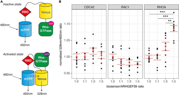
ARHGEF39 was previously implicated in developmental language disorder (DLD) via a functional polymorphism that can disrupt post-transcriptional regulation by microRNAs. ARHGEF39 is part of the family of Rho guanine nucleotide exchange factors (RhoGEFs) that activate small Rho GTPases to regulate a wide variety of cellular processes. However, little is known about the function of ARHGEF39, or how its function might contribute to neurodevelopment or related disorders. Here, we explore the molecular function of ARHGEF39 and show that it activates the Rho GTPase RHOA and that high ARHGEF39 expression in cell cultures leads to an increase of detached cells. To explore its role in neurodevelopment, we analyse published single cell RNA-sequencing data and demonstrate that ARHGEF39 is a marker gene for proliferating neural progenitor cells and that it is co-expressed with genes involved in cell division. This suggests a role for ARHGEF39 in neurogenesis in the developing brain. The co-expression of ARHGEF39 with other RHOA-regulating genes supports RHOA as substrate of ARHGEF39 in neural cells, and the involvement of RHOA in neuropsychiatric disorders highlights a potential link between ARHGEF39 and neurodevelopment and disorder. Understanding the GTPase substrate, co-expression network, and processes downstream of ARHGEF39 provide new avenues for exploring the mechanisms by which altered expression levels of ARHGEF39 may contribute to neurodevelopment and associated disorders.
Keywords: ARHGEF39; RHOA; Rho GTPases; cell adhesion; cell division; neural progenitor cells (NPCs); scRNA-seq.
Copyright © 2022 Anijs, Devanna and Vernes.
Conflict of interest statement
The authors declare that the research was conducted in the absence of any commercial or financial relationships that could be construed as a potential conflict of interest.
Figures


Similar articles
-
ARHGEF39 promotes gastric cancer cell proliferation and migration via Akt signaling pathway.Mol Cell Biochem. 2018 Mar;440(1-2):33-42. doi: 10.1007/s11010-017-3153-3. Epub 2017 Sep 4. Mol Cell Biochem. 2018. PMID: 28871449
-
ARHGEF39 promotes tumor progression via activation of Rac1/P38 MAPK/ATF2 signaling and predicts poor prognosis in non-small cell lung cancer patients.Lab Invest. 2018 May;98(5):670-681. doi: 10.1038/s41374-018-0022-y. Epub 2018 Jan 30. Lab Invest. 2018. PMID: 29382922
-
Rho guanine nucleotide exchange factor 39 increases the viability, migration and invasion of clear cell renal cell carcinoma cells via the activation of the AKT/ERK signaling pathway.Genet Mol Biol. 2020 Nov 18;43(4):e20190383. doi: 10.1590/1678-4685-GMB-2019-0383. eCollection 2020. Genet Mol Biol. 2020. PMID: 33231603 Free PMC article.
-
A current overview of RhoA, RhoB, and RhoC functions in vascular biology and pathology.Biochem Pharmacol. 2022 Dec;206:115321. doi: 10.1016/j.bcp.2022.115321. Epub 2022 Oct 25. Biochem Pharmacol. 2022. PMID: 36306821 Review.
-
MicroRNA Regulation of the Small Rho GTPase Regulators-Complexities and Opportunities in Targeting Cancer Metastasis.Cancers (Basel). 2020 Apr 28;12(5):1092. doi: 10.3390/cancers12051092. Cancers (Basel). 2020. PMID: 32353968 Free PMC article. Review.
Cited by
-
Proteomics Analysis of the Protective Effect of Polydeoxyribonucleotide Extracted from Sea Cucumber (Apostichopus japonicus) Sperm in a Hydrogen Peroxide-Induced RAW264.7 Cell Injury Model.Mar Drugs. 2024 Jul 21;22(7):325. doi: 10.3390/md22070325. Mar Drugs. 2024. PMID: 39057434 Free PMC article.
-
Citalopram exposure of hESCs during neuronal differentiation identifies dysregulated genes involved in neurodevelopment and depression.Front Cell Dev Biol. 2024 Jul 11;12:1428538. doi: 10.3389/fcell.2024.1428538. eCollection 2024. Front Cell Dev Biol. 2024. PMID: 39055655 Free PMC article.
References
Grants and funding
LinkOut - more resources
Full Text Sources
Research Materials
Miscellaneous