

Hybrid surgery for coexistence of cerebral arteriovenous malformation and primitive trigeminal artery: A case report and literature review
- PMID: 35959118
- PMCID: PMC9360567
- DOI: 10.3389/fsurg.2022.888558
Hybrid surgery for coexistence of cerebral arteriovenous malformation and primitive trigeminal artery: A case report and literature review
Abstract
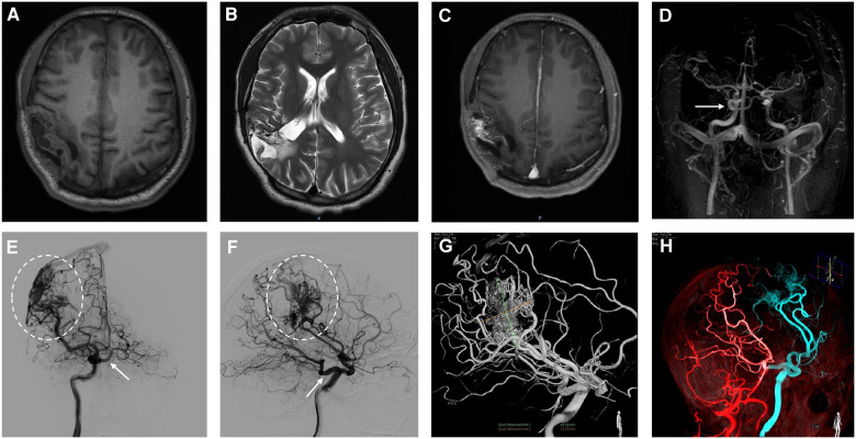
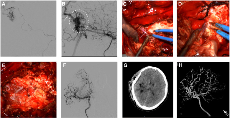
The primitive trigeminal artery (PTA), an abnormal carotid-basilar anastomosis, forms the vascular anomaly connection between the internal carotid artery and vertebrobasilar system. Rarely, PTA can be complicated by several other cerebrovascular disease, including arteriovenous malformations (AVMs), intracranial aneurysms, moyamoya disease, and carotid-cavernous malformations. Herein, we reported a rare case of PTA combined with an AVM in a male patient. The patient was a 28-year-old male with epileptic seizures at the onset of symptoms. Magnetic resonance imaging showed abnormal signal foci and localized softening foci formation with gliosis in the right parietal temporal lobe. Furthermore, using a digital subtraction angiogram (DSA), it was found that an abnormal carotid-basilar anastomosis had developed through a PTA originating from the cavernous portion of the right internal carotid artery (ICA) and a large AVM on the surface of the right carotid artery. The lesion of AVM tightly developed and draining into superior sagittal sinus. A hybrid operating room was used for the surgery. The main feeding arteries of the AVM originating from three major arteries, including the right middle cerebral artery, the right anterior cerebral artery, and the right posterior cerebral artery, were clipped and subsequently, then the AVM was thoroughly removed. The intraoperative DSA showed that the AVM had been resected completely. Postoperative pathological examination of the resected specimen indicated the presence of an AVM. The patient recovered well after surgery and has been symptom-free for more than 3 months. In summary, the pathogenesis of the coexistence of PTA and AVM remains unknown. As highlighted in this case report, hybrid surgery can be used to remove AVMs and can improve the patients' prognosis. To our best knowledge, this is the first case in the literature of successful AVM treatment using hybrid surgery.
Keywords: case report; cerebral arteriovenous malformation; hybrid surgery; literature review; primitive trigeminal artery.
© 2022 Wang, Li, Li, Chai, Chen, Xiong and Yang.
Conflict of interest statement
The authors declare that the research was conducted in the absence of any commercial or financial relationships that could be construed as a potential conflict of interest.
Figures
Similar articles
-
Hybrid surgery for an arteriovenous malformation fed by an accessory middle cerebral artery and drained by a developmental venous anomaly: A case report and literature review.Exp Ther Med. 2018 Sep;16(3):1994-2000. doi: 10.3892/etm.2018.6372. Epub 2018 Jun 29. Exp Ther Med. 2018. PMID: 30186430 Free PMC article.
-
Persistent trigeminal artery associated with an occipital arteriovenous malformation: a case report and literature review.Surg Radiol Anat. 2022 Sep;44(9):1271-1275. doi: 10.1007/s00276-022-03003-9. Epub 2022 Sep 2. Surg Radiol Anat. 2022. PMID: 36056236 Review.
-
Primitive trigeminal artery variant associated with intracranial ruptured aneurysm and cerebral arteriovenous malformation--case report.Neurol Med Chir (Tokyo). 1994 Feb;34(2):104-7. doi: 10.2176/nmc.34.104. Neurol Med Chir (Tokyo). 1994. PMID: 7514756 Review.
-
Internal Carotid Artery Dissecting Aneurysm Associated with Persistent Trigeminal Artery: A Case Report.Curr Med Imaging. 2024;20:1-6. doi: 10.2174/0115734056263907231127170341. Curr Med Imaging. 2024. PMID: 38389372
-
Arteriovenous malformation with an occlusive feeding artery coexisting with unilateral moyamoya disease.J Clin Neurol. 2010 Dec;6(4):216-20. doi: 10.3988/jcn.2010.6.4.216. Epub 2010 Dec 31. J Clin Neurol. 2010. PMID: 21264203 Free PMC article.
Cited by
-
The Relationship between the Permanent Trigeminal Artery and Cerebrovascular Disease: A Meta-Analysis.Iran J Public Health. 2024 May;53(5):988-996. doi: 10.18502/ijph.v53i5.15579. Iran J Public Health. 2024. PMID: 38912150 Free PMC article. Review.
References
Publication types
LinkOut - more resources
Full Text Sources
Research Materials
Miscellaneous