

Case report: Successful multimodal assessment and management of chemothorax
- PMID: 35959134
- PMCID: PMC9360527
- DOI: 10.3389/fsurg.2022.921968
Case report: Successful multimodal assessment and management of chemothorax
Abstract
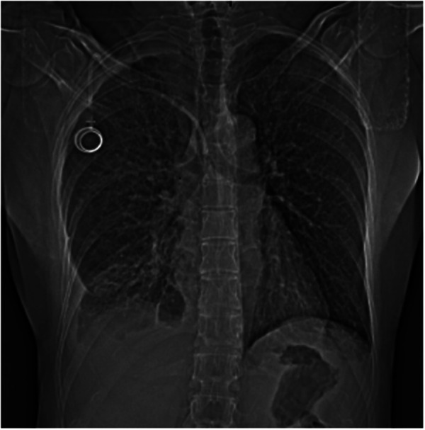
Dislocation or wrong placement of central venous catheters into the pleural cavity is rare, but if undiagnosed, may cause major, sometimes life-threatening, complications (pneumothorax, hemothorax, infection, and migration) and accidental pleural effusion due to intravenous injection of fluids containing drugs (i.e. chemotherapy, antibiotics, parenteral nutrition, other). We report a rare case of pleural effusion consisting of chemotherapy (chemothorax) directly injected into the pleural cavity due to the wrong placement of a central venous catheter (Porth-A-Cath) in a woman with breast cancer. A multidisciplinary management consisting of antidote administration, followed by removal of the venous device and washing of the pleural cavity through video-assisted thoracic surgery (VATS), avoided any major complication related to the adverse event.
Keywords: Port-A-Cath; chemotherapy; chemothorax; dislocation; multidisciplinary management; pleural effusion; thoracoscopy; video-assisted thoracic surgery.
© 2022 Panza, Quercia, Signore, De Iaco, Brascia, Sampietro, Gasbarro, Dell'Aera, Lorusso, Marulli and De Palma.
Conflict of interest statement
The authors declare that the research was conducted in the absence of any commercial or financial relationships that could be construed as a potential conflict of interest.
Figures


Similar articles
-
A delayed complication of a port-a-cath insertion via subclavian venous access: Case report of a "pinch-off syndrome".Int J Surg Case Rep. 2022 May;94:107039. doi: 10.1016/j.ijscr.2022.107039. Epub 2022 Apr 7. Int J Surg Case Rep. 2022. PMID: 35461178 Free PMC article.
-
Thoracoscopy for empyema and hemothorax.Chest. 1996 Jan;109(1):18-24. doi: 10.1378/chest.109.1.18. Chest. 1996. PMID: 8549184
-
Chemothorax: a rare cause of a transudative pleural effusion.BMJ Case Rep. 2015 Dec 10;2015:bcr2015212691. doi: 10.1136/bcr-2015-212691. BMJ Case Rep. 2015. PMID: 26655229 Free PMC article.
-
Practical management of pleural empyema.Monaldi Arch Chest Dis. 2010 Sep;73(3):124-9. doi: 10.4081/monaldi.2010.296. Monaldi Arch Chest Dis. 2010. PMID: 21214042 Review.
-
Complications of central venous port systems: a pictorial review.Insights Imaging. 2019 Aug 28;10(1):86. doi: 10.1186/s13244-019-0770-2. Insights Imaging. 2019. PMID: 31463643 Free PMC article. Review.
Cited by
-
Successful Assessment and Management of Chemothorax: A Case Report and Literature Review.Cureus. 2025 Jun 23;17(6):e86595. doi: 10.7759/cureus.86595. eCollection 2025 Jun. Cureus. 2025. PMID: 40704241 Free PMC article.
References
-
- Medford ARL, Awan YM, Marchbank A, Rahamim J, Unsworth-White J, Pearson PJK. Diagnostic and therapeutic performance of videoassisted thoracoscopic surgery (VATS) in investigation and management of pleural exudates. Ann R Coll Surg Engl. (2008) 90:597–600. 10.1308/003588408X318246 - DOI - PMC - PubMed
Publication types
LinkOut - more resources
Full Text Sources