

Unilateral Multifocal Choroidal Melanoma with Underlying Isolated Choroidal Melanocytosis
- PMID: 35959155
- PMCID: PMC9218616
- DOI: 10.1159/000522262
Unilateral Multifocal Choroidal Melanoma with Underlying Isolated Choroidal Melanocytosis
Abstract
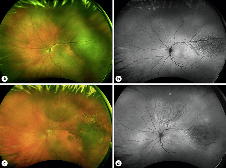
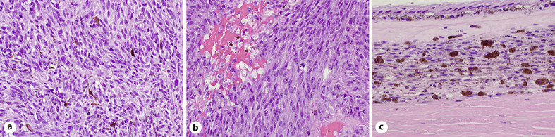
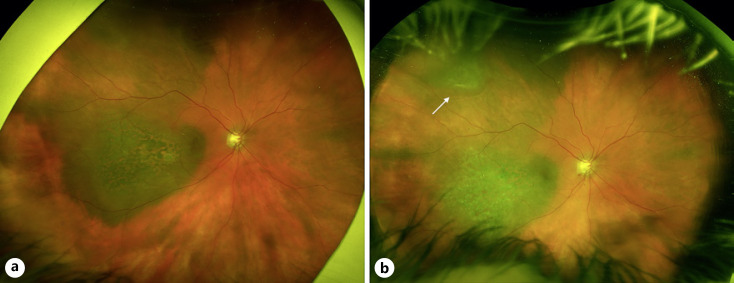
Isolated choroidal melanocytosis is a rare condition that appears to be a limited form of ocular melanocytosis. Ocular melanocytosis has been known to be associated with an increased risk of uveal melanoma, and more recently, a similar association has been suggested for isolated choroidal melanocytosis. We describe 3 cases of patients who developed unilateral, multifocal uveal melanoma in the setting of underlying isolated choroidal melanocytosis. All patients developed either two distinct tumors at presentation or a new discrete choroidal melanoma arising from the choroidal melanocytosis over 1 year following treatment of the original tumor by plaque brachytherapy. These cases provide additional evidence of the association between isolated choroidal melanocytosis and uveal melanoma and suggest increased risk of multifocal melanoma in patients with this condition.
Keywords: Choroidal melanoma; Isolated choroidal melanocytosis; Multifocal choroidal melanoma; Posterior uveal melanoma.
Copyright © 2022 by S. Karger AG, Basel.
Conflict of interest statement
The authors have no conflicts of interest to declare.
Figures
Similar articles
-
Two discrete choroidal melanomas in an eye with ocular melanocytosis.Surv Ophthalmol. 2002 Jan-Feb;47(1):36-41. doi: 10.1016/s0039-6257(01)00281-8. Surv Ophthalmol. 2002. PMID: 11801268 Review.
-
Multifocal Primary Uveal Melanoma: Clinical and Molecular Characteristics.Ocul Oncol Pathol. 2019 Jan;5(1):8-12. doi: 10.1159/000487891. Epub 2018 Apr 27. Ocul Oncol Pathol. 2019. PMID: 30675471 Free PMC article.
-
Intraocular Metastasis in Unilateral Multifocal Uveal Melanoma Without Melanocytosis or Germline BAP1 Mutations.JAMA Ophthalmol. 2019 Dec 1;137(12):1434-1439. doi: 10.1001/jamaophthalmol.2019.3941. JAMA Ophthalmol. 2019. PMID: 31580399 Free PMC article.
-
Isolated choroidal melanocytosis: clinical update on 37 cases.Graefes Arch Clin Exp Ophthalmol. 2020 Dec;258(12):2819-2829. doi: 10.1007/s00417-020-04919-x. Epub 2020 Sep 9. Graefes Arch Clin Exp Ophthalmol. 2020. PMID: 32902757 Free PMC article.
-
Surgical treatment of ocular melanoma.Melanoma Res. 1993 Oct;3(5):305-12. doi: 10.1097/00008390-199310000-00001. Melanoma Res. 1993. PMID: 8292886 Review.
Cited by
-
A case of choroidal melanocytosis observed by multimodal imaging with laser speckle flowgraphy.BMC Ophthalmol. 2023 Apr 26;23(1):180. doi: 10.1186/s12886-023-02933-1. BMC Ophthalmol. 2023. PMID: 37101259 Free PMC article.
References
-
- Teekhasaenee C, Ritch R, Rutnin U, Leelawongs N. Ocular findings in oculodermal melanocytosis. Arch Ophthalmol. 1990;108((8)):1114–20. - PubMed
-
- Singh AD, De Potter P, Fijal BA, Shields CL, Shields JA, Elston RC. Lifetime prevalence of uveal melanoma in white patients with oculo(dermal) melanocytosis. Ophthalmology. 1998;105((1)):195–8. - PubMed
-
- Shields CL, Kaliki S, Livesey M, Walker B, Garoon R, Bucci M, et al. Association of ocular and oculodermal melanocytosis with the rate of uveal melanoma metastasis: analysis of 7872 consecutive eyes. JAMA Ophthalmol. 2013;131((8)):993–1003. - PubMed
-
- Pomeranz GA, Bunt AH, Kalina RE. Multifocal choroidal melanoma in ocular melanocytosis. Arch Ophthalmol. 1981;99((5)):857–63. - PubMed
-
- Honavar SG, Shields CL, Singh AD, Demirci H, Rutledge BK, Shields JA, et al. Two discrete choroidal melanomas in an eye with ocular melanocytosis. Surv Ophthalmol. 2002;47((1)):36–41. - PubMed
Publication types
LinkOut - more resources
Full Text Sources
