

Clinical Applications of Optical Coherence Tomography Angiography in Ocular Oncology: Pearls and Pitfalls
- PMID: 35959157
- PMCID: PMC9218615
- DOI: 10.1159/000520951
Clinical Applications of Optical Coherence Tomography Angiography in Ocular Oncology: Pearls and Pitfalls
Abstract
Background: Optical coherence tomography angiography (OCTA) is a valuable imaging tool for the diagnosis of several retinal and choroidal diseases. Its role in ocular oncology is clinically promising but still controversial. In this review, we report the main applications and limits of the use of OCTA for the study of intraocular tumors.
Summary: OCTA allows a rapid, safe, low-cost, and high-resolution visualization of the retinal and choroidal vasculature. Attempts have been made to use this technology in ocular oncology to differentiate benign and malignant lesions and to assist physicians in the evaluation and monitoring of post-treatment complications. Main limitations include failure in correct segmentation due to the tumor inner profile or thickness, poor penetration of the laser into the lesion, masking effect from overlying fluid, media opacities and poor fixation.
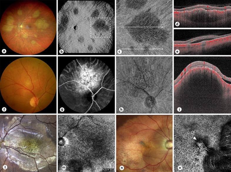
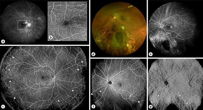
Key messages: The main applications of OCTA in ocular oncology consist of the documentation of tumor-associated choroidal neovascularizations and the study of vascular changes following tumor treatments. In particular, the diffusion of wide-field protocols makes OCTA suitable for the diagnosis and follow-up of radiation chorio-retinopathy, allowing a detailed visualization of both macular and peripheral ischemic changes. Optimistically, future innovations in OCTA technology may offer new perspectives in the diagnosis and follow-up of intraocular tumors.
Keywords: Angiography; Intraocular tumors; Optical coherence tomography angiography; Uveal melanoma.
Copyright © 2021 by S. Karger AG, Basel.
Conflict of interest statement
Giovanni Staurenghi received grants and personal fees from Optovue Inc, Heidelberg Engineering, Zeiss Meditec, Nidek, and CenterVue.
Figures


Similar articles
-
Optical coherence tomography angiography (OCTA) applications in ocular oncology.Eye (Lond). 2020 Sep;34(9):1535-1545. doi: 10.1038/s41433-020-0819-y. Epub 2020 Mar 3. Eye (Lond). 2020. PMID: 32127654 Free PMC article. Review.
-
Retinal Vascular Patterns and Capillary Plexus Reflectivity of Intraocular Tumors; an Optical Coherence Tomography Angiography Study.Curr Eye Res. 2022 Oct;47(10):1424-1435. doi: 10.1080/02713683.2022.2101666. Epub 2022 Jul 25. Curr Eye Res. 2022. PMID: 35819078
-
The application of optical coherence tomography angiography in retinal diseases.Surv Ophthalmol. 2017 Nov-Dec;62(6):838-866. doi: 10.1016/j.survophthal.2017.05.006. Epub 2017 Jun 1. Surv Ophthalmol. 2017. PMID: 28579550 Review.
-
Optical Coherence Tomography Angiography in Retinal Diseases.J Ophthalmic Vis Res. 2016 Jan-Mar;11(1):84-92. doi: 10.4103/2008-322X.180709. J Ophthalmic Vis Res. 2016. PMID: 27195091 Free PMC article. Review.
-
Optical Coherence Tomography Angiography Characteristics of Iris Melanocytic Tumors.Ophthalmology. 2017 Feb;124(2):197-204. doi: 10.1016/j.ophtha.2016.10.003. Epub 2016 Nov 14. Ophthalmology. 2017. PMID: 27856029 Free PMC article.
Cited by
-
Intralesional Vessel Diameter Measured by Optical Coherence Tomography Angiography Could Improve the Differential Diagnosis of Small Melanocytic Choroidal Lesions.Cancers (Basel). 2024 Jun 7;16(12):2167. doi: 10.3390/cancers16122167. Cancers (Basel). 2024. PMID: 38927873 Free PMC article.
References
-
- Shields CL, Pellegrini M, Ferenczy SR, Shields JA. Enhanced depth imaging optical coherence tomography of intraocular tumors: from placid to seasick to rock and rolling topography: the 2013 Francesco Orzalesi Lecture. Retina. 2014;34:1495–512. - PubMed
-
- Spaide RF, Klancnik JM, Jr, Cooney MJ. Retinal vascular layers imaged by fluorescein angiography and optical coherence tomography angiography. JAMA Ophthalmol. 2015;133:45–50. - PubMed
-
- Hirano T, Kakihara S, Toriyama Y, Nittala MG, Murata T, Sadda S. Wide-field en face swept-source optical coherence tomography angiography using extended field imaging in diabetic retinopathy. Br J Ophthalmol. 2017;9:1–5. - PubMed
Publication types
LinkOut - more resources
Full Text Sources
