

Altered functional connectivity within default mode network after rupture of anterior communicating artery aneurysm
- PMID: 35959287
- PMCID: PMC9357996
- DOI: 10.3389/fnagi.2022.905453
Altered functional connectivity within default mode network after rupture of anterior communicating artery aneurysm
Abstract
Background: Rupture of anterior communicating artery (ACoA) aneurysm often leads to cognitive impairment, especially memory complaints. The medial superior frontal gyrus (SFGmed), a node of the default mode network (DMN), has been extensively revealed to participate in various cognitive processes. However, the functional connectivity (FC) characteristics of SFGmed and its relationship with cognitive performance remain unknown after the rupture of the ACoA aneurysm.
Methods: Resting-state functional MRI (fMRI) and cognitive assessment were acquired in 27 eligible patients and 20 controls. Seed-based FC between unilateral SFGmed and the rest of the brain was calculated separately, and then compared their intensity differences between the two groups. Furthermore, we analyzed the correlation between abnormal FC and cognitive function in patients with ruptured ACoA aneurysm.
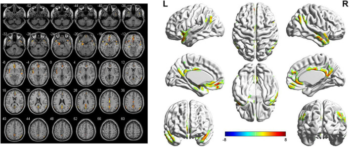
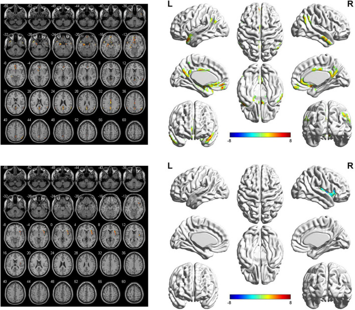
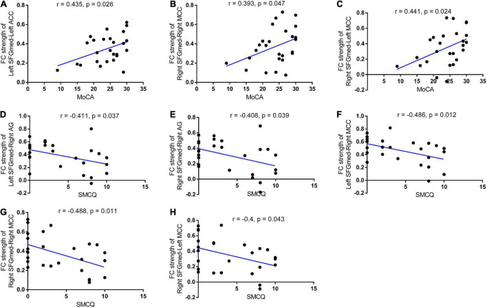
Results: Cognitive impairment was confirmed in 51.9% of the patients. Compared with the controls, patients suffering from ruptured ACoA aneurysm exhibited a similar FC decline between each side of SFGmed and predominant nodes within DMN, including the precuneus, angular gyrus, cingulate cortex, left hippocampus, left amygdala, left temporal pole (TPO), and left medial orbitofrontal cortex (mOFC). Besides, significantly decreased FC of left SFGmed and left insula, right middle temporal gyrus (MTG), as well as right mOFC, were also found. In addition, only enhanced insular connectivity with right SFGmed was determined, whereas increased FC of the left SFGmed was not observed. Correlation analyses showed that lower total cognitive performance or stronger subjective memory complaints were related to reduced connectivity in the SFGmed and several cortical regions such as the angular gyrus and middle cingulate cortex (MCC).
Conclusion: Our results suggest that patients with ruptured ACoA aneurysm exist long-term cognitive impairment and intrinsic hypoconnectivity of cognition-related brain regions within DMN. Deactivation of DMN may be a potential neural mechanism leading to cognitive deficits in these patients.
Keywords: anterior communicating artery aneurysm; cognitive impairment; default mode network; functional connectivity; resting-state fMRI; subarachnoid hemorrhage.
Copyright © 2022 Chen, Kang, Yu, Lin, Dai, Yu, Wang, Sun and Kang.
Conflict of interest statement
The authors declare that the research was conducted in the absence of any commercial or financial relationships that could be construed as a potential conflict of interest.
Figures
Similar articles
-
Altered hippocampal functional connectivity after the rupture of anterior communicating artery aneurysm.Front Aging Neurosci. 2022 Nov 7;14:997231. doi: 10.3389/fnagi.2022.997231. eCollection 2022. Front Aging Neurosci. 2022. PMID: 36420312 Free PMC article.
-
Dysfunction of the Default Mode Network in Drug-Naïve Parkinson's Disease with Mild Cognitive Impairments: A Resting-State fMRI Study.Front Aging Neurosci. 2016 Oct 26;8:247. doi: 10.3389/fnagi.2016.00247. eCollection 2016. Front Aging Neurosci. 2016. PMID: 27833548 Free PMC article.
-
Patterns of default mode network in temporal lobe epilepsy with and without hippocampal sclerosis.Epilepsy Behav. 2021 Aug;121(Pt B):106523. doi: 10.1016/j.yebeh.2019.106523. Epub 2019 Oct 20. Epilepsy Behav. 2021. PMID: 31645315
-
Dysregulation within the salience network and default mode network in hyperthyroid patients: a follow-up resting-state functional MRI study.Brain Imaging Behav. 2020 Feb;14(1):30-41. doi: 10.1007/s11682-018-9961-6. Brain Imaging Behav. 2020. PMID: 30259292 Review.
-
Rectus gyrus hematoma: An overview.Surg Neurol Int. 2022 Dec 2;13:558. doi: 10.25259/SNI_1023_2022. eCollection 2022. Surg Neurol Int. 2022. PMID: 36600763 Free PMC article. Review.
Cited by
-
Neurobehavioral impairments predict specific cerebral damage in rat model of subarachnoid hemorrhage.Res Sq [Preprint]. 2023 May 19:rs.3.rs-2943917. doi: 10.21203/rs.3.rs-2943917/v1. Res Sq. 2023. Update in: Transl Stroke Res. 2024 Oct;15(5):950-969. doi: 10.1007/s12975-023-01180-2. PMID: 37292945 Free PMC article. Updated. Preprint.
-
Neurobehavioral Impairments Predict Specific Cerebral Damage in Rat Model of Subarachnoid Hemorrhage.Transl Stroke Res. 2024 Oct;15(5):950-969. doi: 10.1007/s12975-023-01180-2. Epub 2023 Jul 26. Transl Stroke Res. 2024. PMID: 37493939
References
-
- Beeckmans K., Crunelle C., Van den Bossche J., Dierckx E., Michiels K., Vancoillie P., et al. (2020). Cognitive outcome after surgical clipping versus endovascular coiling in patients with subarachnoid hemorrhage due to ruptured anterior communicating artery aneurysm. Acta Neurol. Belg. 120 123–132. - PubMed
LinkOut - more resources
Full Text Sources