Clinicoradiographic evaluation of advanced-platelet rich fibrin block (A PRF + i PRF + nanohydroxyapatite) compared to nanohydroxyapatite alone in the management of periodontal intrabony defects
- PMID: 35959304
- PMCID: PMC9362812
- DOI: 10.4103/jisp.jisp_882_20
Clinicoradiographic evaluation of advanced-platelet rich fibrin block (A PRF + i PRF + nanohydroxyapatite) compared to nanohydroxyapatite alone in the management of periodontal intrabony defects
Abstract
Background: Several bone grafting formulations have been given clinically acceptable outcomes in treating intrabony defects. Platelet rich fibrin (PRF), an autologous platelet concentrate holds potential to be used for regenerative treatment. The purpose of this study was to evaluate clinical and radiographic outcomes in periodontal intrabony defects treated with advanced-PRF block (A PRF + i PRF + nanohydroxyapatite [nHA]) compared to nHA alone.
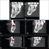
Methods: Twenty-eight sites in chronic periodontitis patients having probing pocket depth (PPD) ≥6 mm and 3 walled intrabony defects (depth of ≥3 mm) were selected, randomly allotted into two groups: Group A was treated with A-PRF block and Group B with nHA (Sybograf™). Clinical parameters including plaque index (PI), gingival index (GI), PPD, relative attachment level (RAL) and radiographically linear and volumetric defect fill were assessed using cone beam computed tomography at baseline and 6 months postoperatively.
Results: Intragroup comparison using paired t-test and intergroup comparison using unpaired t-test was done. Group A demonstrated significantly higher reduction in PPD and gain in RAL when compared to Group B (P ≤ 0.05) at the end of 6 months. Similarly gain in bone volume was greater in Group A (0.1 ± 0.05) as compared to Group B (0.04 ± 0.02) (P ≤ 0.05).
Conclusion: Advanced-PRF block showed significant clinical and radiographic improvement as compared to nHA alone which depicts that, it may be an ideal graft to be used for the treatment of periodontal intrabony defects.
Keywords: Intrabony defects; nanohydroxyapatite; periodontal regeneration; platelet rich fibrin.
Copyright: © 2022 Indian Society of Periodontology.
Conflict of interest statement
There are no conflicts of interest.
Figures


Similar articles
-
Comparative evaluation of regenerative potential of injectable platelet-rich fibrin and platelet-rich fibrin with demineralized freeze-dried bone allograft in the treatment of intrabony defects: A randomized controlled clinical study.Natl J Maxillofac Surg. 2023 Sep-Dec;14(3):399-405. doi: 10.4103/njms.njms_39_22. Epub 2023 Nov 10. Natl J Maxillofac Surg. 2023. PMID: 38273925 Free PMC article.
-
Injectable platelet-rich fibrin with demineralized freeze-dried bone allograft compared to demineralized freeze-dried bone allograft in intrabony defects of patients with stage-III periodontitis: a randomized controlled clinical trial.Clin Oral Investig. 2023 Jul;27(7):3457-3467. doi: 10.1007/s00784-023-04954-y. Epub 2023 Mar 31. Clin Oral Investig. 2023. PMID: 37002441 Free PMC article. Clinical Trial.
-
Clinical and radiographic evaluation of platelet-rich fibrin as an adjunct to bone grafting demineralized freeze-dried bone allograft in intrabony defects.J Indian Soc Periodontol. 2020 Jan-Feb;24(1):60-66. doi: 10.4103/jisp.jisp_99_19. Epub 2020 Jan 2. J Indian Soc Periodontol. 2020. PMID: 31983847 Free PMC article.
-
Use of Platelet-Rich Fibrin in the Treatment of Periodontal Intrabony Defects: A Systematic Review and Meta-Analysis.Biomed Res Int. 2021 Feb 4;2021:6669168. doi: 10.1155/2021/6669168. eCollection 2021. Biomed Res Int. 2021. Retraction in: Biomed Res Int. 2024 Mar 20;2024:9838732. doi: 10.1155/2024/9838732. PMID: 33614786 Free PMC article. Retracted.
-
Actual quantitative attachment gain secondary to use of autologous platelet concentrates in the treatment of intrabony defects: A meta-analysis.J Indian Soc Periodontol. 2019 May-Jun;23(3):190-202. doi: 10.4103/jisp.jisp_498_18. J Indian Soc Periodontol. 2019. PMID: 31142999 Free PMC article. Review.
Cited by
-
Influence of alloplastic materials, biologics, and their combinations, along with defect characteristics, on short-term intrabony defect surgical treatment outcomes: a systematic review and network meta-analysis.BMC Oral Health. 2025 Mar 20;25(1):413. doi: 10.1186/s12903-025-05782-0. BMC Oral Health. 2025. PMID: 40114125 Free PMC article.
-
Alveolar Bone Preservation Using a Combination of Nanocrystalline Hydroxyapatite and Injectable Platelet-Rich Fibrin: A Study in Rats.Curr Issues Mol Biol. 2023 Jul 17;45(7):5967-5980. doi: 10.3390/cimb45070377. Curr Issues Mol Biol. 2023. PMID: 37504293 Free PMC article.
References
-
- Karring T, Nyman S, Gottlow J, Laurell L. Development of the biological concept of guided tissue regeneration – Animal and human studies. Periodontol 2000. 1993;1:26–35. - PubMed
-
- Ivanovic A, Nikou G, Miron RJ, Nikolidakis D, Sculean A. Which biomaterials may promote periodontal regeneration in intrabony periodontal defects? A systematic review of preclinical studies. Quintessence Int. 2014;45:385–95. - PubMed
-
- Laurencin C, Khan Y, El-Amin SF. Bone graft substitutes. Expert Rev Med Devices. 2006;3:49–57. - PubMed
-
- Murugan R, Ramakrishna S. Development of nanocomposites for bone grafting. Comp Sci Tech. 2005;65:2385–406.
LinkOut - more resources
Full Text Sources
Research Materials
Miscellaneous
