Effects of antibiotic treatment and phagocyte infiltration on development of Pseudomonas aeruginosa biofilm-Insights from the application of a novel PF hydrogel model in vitro and in vivo
- PMID: 35959369
- PMCID: PMC9362844
- DOI: 10.3389/fcimb.2022.826450
Effects of antibiotic treatment and phagocyte infiltration on development of Pseudomonas aeruginosa biofilm-Insights from the application of a novel PF hydrogel model in vitro and in vivo
Abstract
Background and purpose: Bacterial biofilm infections are major health issues as the infections are highly tolerant to antibiotics and host immune defenses. Appropriate biofilm models are important to develop and improve to make progress in future biofilm research. Here, we investigated the ability of PF hydrogel material to facilitate the development and study of Pseudomonas aeruginosa biofilms in vitro and in vivo.
Methods: Wild-type P. aeruginosa PAO1 bacteria were embedded in PF hydrogel situated in vitro or in vivo, and the following aspects were investigated: 1) biofilm development; 2) host immune response and its effect on the bacteria; and 3) efficacy of antibiotic treatment.
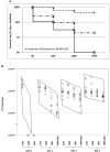
Results: Microscopy demonstrated that P. aeruginosa developed typical biofilms inside the PF hydrogels in vitro and in mouse peritoneal cavities where the PF hydrogels were infiltrated excessively by polymorphonuclear leukocytes (PMNs). The bacteria remained at a level of ~106 colony-forming unit (CFU)/hydrogel for 7 days, indicating that the PMNs could not eradicate the biofilm bacteria. β-Lactam or aminoglycoside mono treatment at 64× minimal inhibitory concentration (MIC) killed all bacteria in day 0 in vitro biofilms, but not in day 1 and older biofilms, even at a concentration of 256× MIC. Combination treatment with the antibiotics at 256× MIC completely killed the bacteria in day 1 in vitro biofilms, and combination treatment in most of the cases showed significantly better bactericidal effects than monotherapies. However, in the case of the established in vivo biofilms, the mono and combination antibiotic treatments did not efficiently kill the bacteria.
Conclusion: Our results indicate that the bacteria formed typical biofilms in PF hydrogel in vitro and in vivo and that the biofilm bacteria were tolerant against antibiotics and host immunity. The PF hydrogel biofilm model is simple and easy to fabricate and highly reproducible with various application possibilities. We conclude that the PF hydrogel biofilm model is a new platform that will facilitate progress in future biofilm investigations, as well as studies of the efficacy of new potential medicine against biofilm infections.
Keywords: PF hydrogel; Pseudomonas aeruginosa; antibiotic resistance; biofilm infection; in vivo model.
Copyright © 2022 Wu, Song, Yam, Plotkin, Wang, Rybtke, Seliktar, Kofidis, Høiby, Tolker-Nielsen, Song and Givskov.
Conflict of interest statement
The authors declare that the research was conducted in the absence of any commercial or financial relationships that could be construed as a potential conflict of interest.
Figures


Similar articles
-
The Efficacy of Colistin Combined with Amikacin or Levofloxacin against Pseudomonas aeruginosa Biofilm Infection.Microbiol Spectr. 2022 Oct 26;10(5):e0146822. doi: 10.1128/spectrum.01468-22. Epub 2022 Sep 14. Microbiol Spectr. 2022. PMID: 36102678 Free PMC article.
-
Baicalin inhibits biofilm formation, attenuates the quorum sensing-controlled virulence and enhances Pseudomonas aeruginosa clearance in a mouse peritoneal implant infection model.PLoS One. 2017 Apr 28;12(4):e0176883. doi: 10.1371/journal.pone.0176883. eCollection 2017. PLoS One. 2017. PMID: 28453568 Free PMC article.
-
Antibiotics Enhance Prevention and Eradication Efficacy of Cathodic-Voltage-Controlled Electrical Stimulation against Titanium-Associated Methicillin-Resistant Staphylococcus aureus and Pseudomonas aeruginosa Biofilms.mSphere. 2019 May 1;4(3):e00178-19. doi: 10.1128/mSphere.00178-19. mSphere. 2019. PMID: 31043516 Free PMC article.
-
Pseudomonas aeruginosa biofilm infections: from molecular biofilm biology to new treatment possibilities.APMIS Suppl. 2014 Dec;(138):1-51. doi: 10.1111/apm.12335. APMIS Suppl. 2014. PMID: 25399808 Review.
-
Pseudomonas aeruginosa chromosomal beta-lactamase in patients with cystic fibrosis and chronic lung infection. Mechanism of antibiotic resistance and target of the humoral immune response.APMIS Suppl. 2003;(116):1-47. APMIS Suppl. 2003. PMID: 14692154 Review.
Cited by
-
Comparative in vitro activity of various antibiotic against planktonic and biofilm and the gene expression profile in Pseudomonas aeruginosa.AIMS Microbiol. 2023 Mar 30;9(2):313-331. doi: 10.3934/microbiol.2023017. eCollection 2023. AIMS Microbiol. 2023. PMID: 37091817 Free PMC article.
-
In vivo evolution of antimicrobial resistance in a biofilm model of Pseudomonas aeruginosa lung infection.ISME J. 2024 Jan 8;18(1):wrae036. doi: 10.1093/ismejo/wrae036. ISME J. 2024. PMID: 38478426 Free PMC article.
References
-
- Aaron S. D., Ferris W., Ramotar K., Vandemheen K., Chan F., Saginur R. (2002). Single and combination antibiotic susceptibilities of planktonic, adherent, and biofilm-grown Pseudomonas aeruginosa isolates cultured from sputa of adults with cystic fibrosis. J. Clin. Microbiol. 40, 4172–4179. doi: 10.1128/jcm.40.11.4172-4179.2002 - DOI - PMC - PubMed
-
- Christensen L. D., Moser C., Jensen P. O., Rasmussen T. B., Christophersen L., Kjelleberg S., et al. . (2007). Impact of Pseudomonas aeruginosa quorum sensing on biofilm persistence in an in vivo intraperitoneal foreign-body infection model. Microbiology. 153, 2312–2320. doi: 10.1099/mic.0.2007/006122-0 - DOI - PubMed
-
- Clark D. J., Maaløe O. (1967). DNA Replication and the division cycle in Escherichia coli . J. Mol. Biol. 23, 99–112. doi: 10.1016/S0022-2836(67)80070-6 - DOI
Publication types
MeSH terms
Substances
LinkOut - more resources
Full Text Sources

