

Organization of the ventricular zone of the cerebellum
- PMID: 35959470
- PMCID: PMC9358289
- DOI: 10.3389/fncel.2022.955550
Organization of the ventricular zone of the cerebellum
Abstract
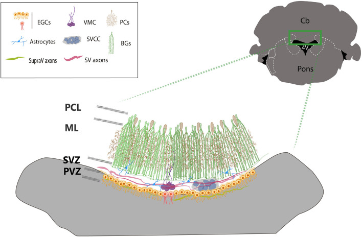
The roof of the fourth ventricle (4V) is located on the ventral part of the cerebellum, a region with abundant vascularization and cell heterogeneity that includes tanycyte-like cells that define a peculiar glial niche known as ventromedial cord. This cord is composed of a group of biciliated cells that run along the midline, contacting the ventricular lumen and the subventricular zone. Although the complex morphology of the glial cells composing the cord resembles to tanycytes, cells which are known for its proliferative capacity, scarce or non-proliferative activity has been evidenced in this area. The subventricular zone of the cerebellum includes astrocytes, oligodendrocytes, and neurons whose function has not been extensively studied. This review describes to some extent the phenotypic, morphological, and functional characteristics of the cells that integrate the roof of the 4V, primarily from rodent brains.
Keywords: Bergmann glia; astrocytes; choroid plexus; ependymal glial cells; fourth ventricle.
Copyright © 2022 Gómez-González, Becerra-González, Martínez-Mendoza, Rodríguez-Arzate and Martínez-Torres.
Figures
Similar articles
-
Response to Hypoxic Preconditioning of Glial Cells from the Roof of the Fourth Ventricle.Neuroscience. 2020 Jul 15;439:211-229. doi: 10.1016/j.neuroscience.2019.09.015. Epub 2019 Nov 2. Neuroscience. 2020. PMID: 31689390
-
Identification of novel cellular clusters define a specialized area in the cerebellar periventricular zone.Sci Rep. 2017 Jan 20;7:40768. doi: 10.1038/srep40768. Sci Rep. 2017. PMID: 28106069 Free PMC article.
-
Inter-fastigial projections along the roof of the fourth ventricle.Brain Struct Funct. 2021 Apr;226(3):901-917. doi: 10.1007/s00429-021-02217-8. Epub 2021 Jan 28. Brain Struct Funct. 2021. PMID: 33511462
-
Origin, lineage and function of cerebellar glia.Prog Neurobiol. 2013 Oct;109:42-63. doi: 10.1016/j.pneurobio.2013.08.001. Epub 2013 Aug 25. Prog Neurobiol. 2013. PMID: 23981535 Review.
-
Cytodifferentiation of Bergmann glia and its relationship with Purkinje cells.Anat Sci Int. 2002 Jun;77(2):94-108. doi: 10.1046/j.0022-7722.2002.00021.x. Anat Sci Int. 2002. PMID: 12418089 Review.
References
-
- Albors A. R., Singer G. A., May A. P., Ponting C. P., Storey K. G. (2022). Ependymal cell maturation is heterogeneous and ongoing in the mouse spinal cord and dynamically regulated in response to injury. bioRxiv [Preprint]. 10.1101/2022.03.07.483249 - DOI
-
- Altman J., Bayer S. A. (1997). Development of the Cerebellar System in Relation to Its Evolution, Structure and Functions. Boca Raton, FL: CRC Press.
-
- Alvarez-Morujo A. J., Toranzo D., Blázquez J. L., Peláez B., Sánchez A., Pastor F. E., et al. . (1992). The ependymal surface of the fourth ventricle of the rat: a combined scanning and transmission electron microscopic study. Histol. Histopathol. 7, 259–266. - PubMed
Publication types
LinkOut - more resources
Full Text Sources