Review
. 2022 Jul 26:10:969547.
doi: 10.3389/fcell.2022.969547. eCollection 2022.

Amyloid precursor protein (APP) and amyloid β (Aβ) interact with cell adhesion molecules: Implications in Alzheimer's disease and normal physiology

Affiliations
Review

Amyloid precursor protein (APP) and amyloid β (Aβ) interact with cell adhesion molecules: Implications in Alzheimer's disease and normal physiology

Grant Pfundstein et al. Front Cell Dev Biol. .

Abstract

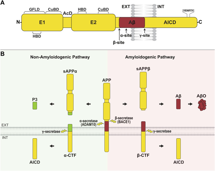
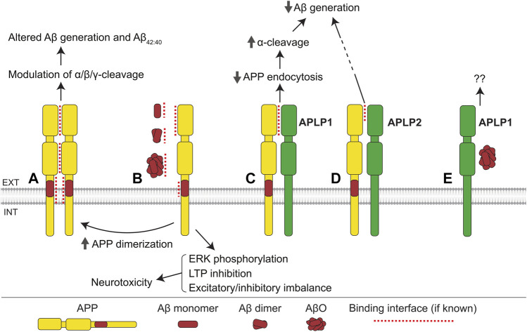
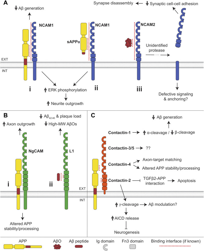
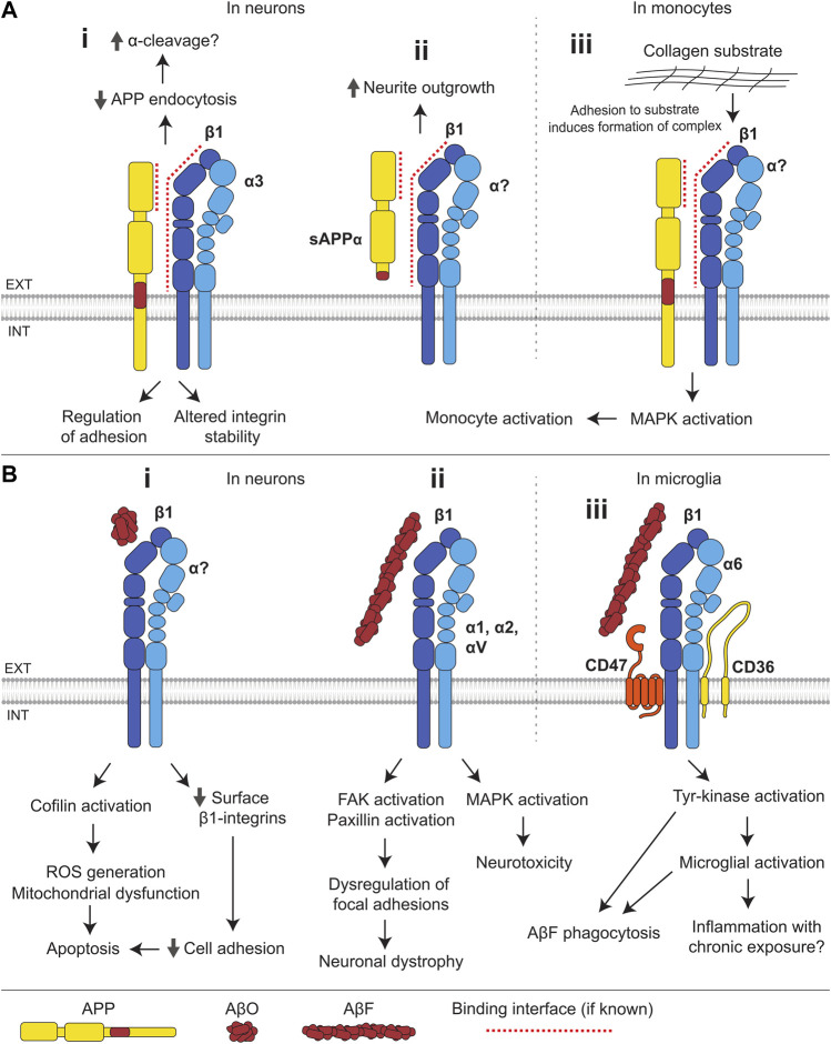
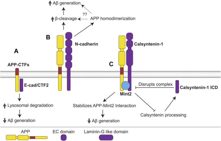
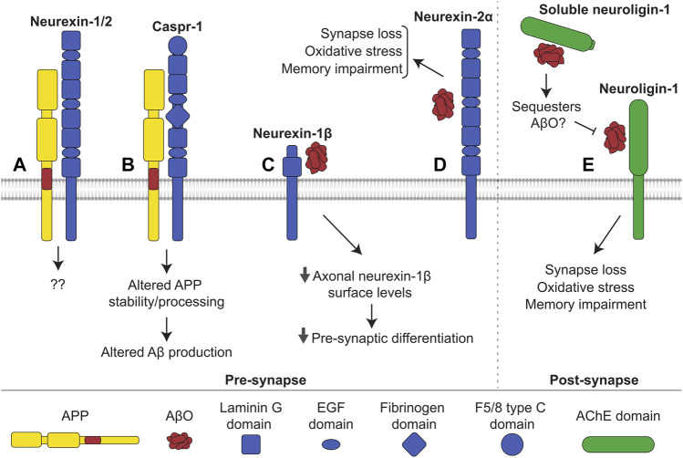
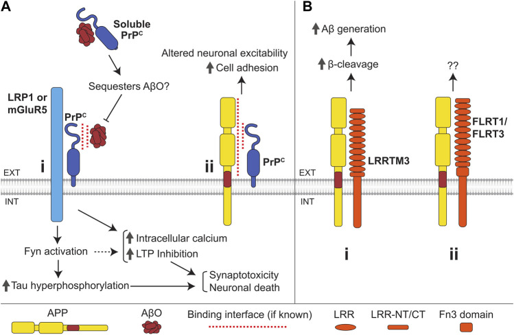
Alzheimer's disease (AD) is an incurable neurodegenerative disorder in which dysfunction and loss of synapses and neurons lead to cognitive impairment and death. Accumulation and aggregation of neurotoxic amyloid-β (Aβ) peptides generated via amyloidogenic processing of amyloid precursor protein (APP) is considered to play a central role in the disease etiology. APP interacts with cell adhesion molecules, which influence the normal physiological functions of APP, its amyloidogenic and non-amyloidogenic processing, and formation of Aβ aggregates. These cell surface glycoproteins also mediate attachment of Aβ to the neuronal cell surface and induce intracellular signaling contributing to Aβ toxicity. In this review, we discuss the current knowledge surrounding the interactions of cell adhesion molecules with APP and Aβ and analyze the evidence of the critical role these proteins play in regulating the processing and physiological function of APP as well as Aβ toxicity. This is a necessary piece of the complex AD puzzle, which we should understand in order to develop safe and effective therapeutic interventions for AD.

Keywords: Alzheimer’s disease; amyloid precursor protein (APP); amyloid-beta; cell adhesion molecule (CAM); immunoglobulin superfamily; integrin; neurexin; prion protein (PrP).


Conflict of interest statement

The authors declare that the research was conducted in the absence of any commercial or financial relationships that could be construed as a potential conflict of interest.

Figures

FIGURE 1
FIGURE 1
APP structure and processing. (A) APP is composed of a large N-terminal extracellular domain, transmembrane region, and short cytoplasmic tail. The extracellular domain comprises two rigidly folded regions, E1 and E2, joined by an acidic domain (AcD). E1 contains a heparin-binding domain (HBD) within a larger growth factor-like domain (GFLD), and a copper/zinc-binding domain (CuBD). E2 comprises the second HBD and CuBD. The juxtamembrane region contains the α- and β-cleavage sites, while the γ-cleavage site is located within the transmembrane domain. The C-terminal intracellular domain (AICD) contains the YENPTY sequence, which binds cytosolic adaptor proteins. (B) APP is primarily processed along two opposing pathways. In amyloidogenic processing, APP is cleaved by β-secretase (BACE1) at the N-terminus of Aβ, generating sAPPβ and the membrane-bound β-CTF. Subsequent γ-secretase cleavage of β-CTF releases the Aβ peptide into the extracellular/lumenal space and AICD into the cytosol. Aβ peptides aggregate and form oligomers (AβO). In non-amyloidogenic processing, APP is cleaved by α-secretase (ADAM10) within the Aβ region, producing sAPPα and α-CTF. Ensuing cleavage of α-CTF by γ-secretase liberates P3 into the extracellular/lumenal space and AICD into the cytosol.
FIGURE 2
FIGURE 2
APP family interactions. (A) Interactions between E1, E2, and transmembrane domains of APP mediate formation of homodimers. Homodimerization influences the α-, β-, and γ-cleavage of APP, Aβ generation, and the ratio of Aβ42/40. (B) Monomers, dimers, and oligomers of Aβ bind to the E1 domain and cognate Aβ region of APP. The interaction with Aβ monomers and dimers promotes APP homodimerization, while the APP-AβO interaction induces ERK phosphorylation, inhibits long-term potentiation (LTP), and leads to excitatory/inhibitory imbalance, ultimately resulting in neurotoxicity. (C) APLP1 interacts with the E1 domain of APP, suppressing APP endocytosis, increasing cell surface levels of APP, and thereby facilitating the α-cleavage of APP, and consequently reducing Aβ generation. (D) APLP2 interacts with APP and reduces Aβ generation via an unknown mechanism. (E) APLP1 interacts with AβOs with unknown consequence.
FIGURE 3
FIGURE 3
Interactions of IgSF CAMs with APP and Aβ. (A) (i) NCAM1 interacts with the E2 domain of APP and reduces Aβ generation. Binding of APP or (ii) sAPPα to NCAM1 induces ERK phosphorylation and promotes neurite outgrowth. (iii) AβOs bind to NCAM2 triggering shedding of its extracellular domain by an unidentified protease, causing synapse disassembly. (B) (i) The chicken ortholog of L1, NgCAM, interacts with APP and regulates APP stability and processing. Binding of APP to NgCAM promotes axon outgrowth in retinal ganglion cells. (ii) Aβ peptides bind to the second Fn3 domain of L1. L1 reduces Aβ42:40 ratio and the formation of high molecular-weight (MW) AβOs in favor of monomers, dimers, and tetramers. (C) APP interacts with all contactin family members except for contactin-6. The E1 CuBD of APP binds to the second Fn3 domain of contactin. Contactin-1 promotes α-cleavage and reduces β-cleavage of APP, thereby reducing Aβ generation. Contactin-2 promotes γ-cleavage of APP, thereby increasing AICD release and inhibiting neurogenesis. Contactin-2 also inhibits the binding of APP to TGFβ2 and reduces apoptosis induced by APP-TGFβ2 interactions. Contactin-4 regulates APP stability and processing and is required for APP-dependent axon-target matching. The function of APP interactions with contactin-3 and -5 remains undetermined.
FIGURE 4
FIGURE 4
Interactions of integrins with APP and Aβ. (A) (i) The E1 domain of APP interacts with β1-and α3-integrins. Integrins suppress endocytosis of APP and thereby may facilitate its α-cleavage. APP influences integrin stability and integrin-dependent adhesion. (ii) In neurons, sAPPα binds to β1-integrins and induces neurite outgrowth. (iii) In monocytes, adhesion to a collagen substrate induces the formation of an APP-β1-integrin complex, which activates MAPK signaling resulting in monocyte activation. (B) (i) In neurons, binding of AβOs to β1-integrins leads to a reduction in the cell surface levels of β1-integrins and compromised cell adhesion, and results in activation of the f-actin severing protein, cofilin, mitochondrial dysfunction and ROS generation, leading to apoptosis. (ii) AβFs bind several integrin subunits (β1, α1, α2, and αV) and induce MAPK activation and neurotoxicity. Binding of AβF to β1-integrin also induces FAK and paxillin activation, leading to dysregulation of integrin-mediated focal adhesion and contributing to neuronal dystrophy. (iii) In microglia, AβFs bind to a receptor complex composed of α6β1-integrin, CD47, and CD36, and induce tyrosine-kinase activation, stimulating AβF phagocytosis and microglial activation.
FIGURE 5
FIGURE 5
Interactions of cadherins with APP. (A) The γ-cleavage product of E-cadherin, E-cad/CTF2, interacts with APP-CTFs, promoting their lysosomal degradation and precluding Aβ generation. (B) N-cadherin interacts with APP promoting its homodimerization, β-cleavage, and Aβ generation. N-cadherin-induced APP homodimerization may promote β-cleavage and Aβ generation, however, this mechanism needs to be confirmed. (C) APP forms a complex with the non-classical cadherin, calsyntenin-1, and adaptor protein Mint2. The APP-Mint2 interaction, which may suppress Aβ generation, is stabilized in the tripartite complex with calsyntenin-1, thus enabling greater suppression of Aβ generation. Formation of the APP-Mint2-calsyntenin-1 complex also precludes calsyntenin-1 processing, reducing generation of the γ-cleavage product, calsyntenin-1 ICD, which disrupts the APP-Mint2-calsyntenin-1 complex.
FIGURE 6
FIGURE 6
Interactions of neurexin and neuroligin with APP and Aβ. (A) Neurexin-1 and -2 interact with APP in mouse brains, with unknown consequence. (B) Caspr-1 interacts with APP. This interaction alters stability and processing of APP influencing production of Aβ. It is uncertain whether the interaction increases or decreases amyloidogenic processing. (C) AβOs bind to neurexin-1β, reducing its levels at the axonal cell surface and hindering presynaptic differentiation. (D) AβOs additionally bind to neurexin-2α triggering oxidative stress, synapse loss, and memory impairments in mice. Similarly, (E) AβOs interact with neuroligin-1 at the post-synapse, inducing oxidative stress, synapse loss, and memory impairments in mice. Shedding releases soluble neuroligin-1 which also binds to and likely sequesters AβOs, preventing interactions with membrane-bound neuroligin-1 and other receptors.
FIGURE 7
FIGURE 7
Interactions of the cellular prion protein (PrPc) and LRR superfamily CAMs with APP and Aβ. (A) (i) Binding of AβOs to PrPc induces an increase in intracellular Ca2+ levels, LTP inhibition, Fyn activation, and tau hyperphosphorylation, ultimately resulting in synaptotoxicity and neuronal death. PrPc is anchored to the outer leaflet of the plasma membrane and transmits the AβO-induced signals across the plasma membrane by interacting with the transmembrane proteins LRP1 and mGluR5. Soluble PrPc binds to and can sequester AβOs, preventing membrane-bound PrPc-mediated AβO toxicity. (ii) Interaction of APP with the N-terminal domain of PrPc enhances cell adhesion via an unknown mechanism and may also regulate neuronal excitability. (B) (i) LRRTM3 interacts with APP promoting its β-cleavage and facilitating Aβ generation. (ii) APP also interacts with FLRT1 and FLRT3, with unknown consequence.

Similar articles

Cited by

References

    1. Agiostratidou G., Muros R. M., Shioi J., Marambaud P., Robakis N. K. (2006). The cytoplasmic sequence of E-cadherin promotes non-amyloidogenic degradation of Aβ precursors. J. Neurochem. 96, 1182–1188. 10.1111/j.1471-4159.2005.03616.x - DOI - PubMed
    1. Aisa B., Gil-Bea F. J., Solas M., García-Alloza M., Chen C. P., Lai M. K., et al. (2010). Altered NCAM expression associated with the cholinergic system in Alzheimer's disease. J. Alzheimers Dis. 20, 659–668. 10.3233/JAD-2010-1398 - DOI - PubMed
    1. Alzheimer’s Association (2022). 2022 Alzheimer's disease facts and figures. Alzheimer's Dementia 18, 700–789. - PubMed
    1. Anderson K. L., Ferreira A. (2004). alpha1 integrin activation: a link between beta-amyloid deposition and neuronal death in aging hippocampal neurons. J. Neurosci. Res. 75, 688–697. 10.1002/jnr.20018 - DOI - PubMed
    1. Andrew R. J., Kellett K. A. B., Thinakaran G., Hooper N. M. (2016). A Greek tragedy: The growing complexity of alzheimer amyloid precursor protein proteolysis. J. Biol. Chem. 291, 19235–19244. 10.1074/jbc.R116.746032 - DOI - PMC - PubMed