. 2022 Aug 5;3(3):101606.
doi: 10.1016/j.xpro.2022.101606. eCollection 2022 Sep 16.

Characterizing single-molecule dynamics of viral RNA-dependent RNA polymerases with multiplexed magnetic tweezers

Affiliations

Characterizing single-molecule dynamics of viral RNA-dependent RNA polymerases with multiplexed magnetic tweezers

Louis Kuijpers et al. STAR Protoc. .

Abstract

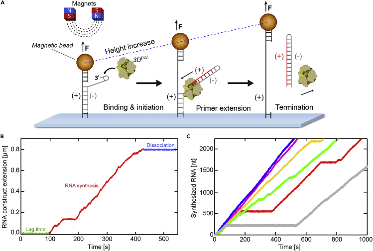
Multiplexed single-molecule magnetic tweezers (MT) have recently been employed to probe the RNA synthesis dynamics of RNA-dependent RNA polymerases (RdRp). Here, we present a protocol for simultaneously probing the RNA synthesis dynamics of hundreds of single polymerases with MT. We describe the preparation of a dsRNA construct for probing single RdRp kinetics. We then detail the measurement of RdRp RNA synthesis kinetics using MT. The protocol is suitable for high-throughput probing of RdRp-targeting antiviral compounds for mechanistic function and efficacy. For complete details on the use and execution of this protocol, please refer to Janissen et al. (2021).

Keywords: Biophysics; Microbiology; Molecular biology; Molecular/Chemical probes; Single-molecule assays.


Conflict of interest statement

The authors declare no competing interests.

Figures

None
Graphical abstract
Figure 1
Figure 1
dsRNA construct preparation for the RdRp RNA synthesis assay (A) Simplified pBAD plasmid maps for PCR amplification of the five DNA fragments. (B) Annealing of the RNA transcripts to generate the dsRNA construct used in MT. The transcripts originate from run-off T7 transcription of the individual DNA fragments. BIO: biotin-enriched handle; DIG: digoxigenin-enriched handle; SP: spacer fragment; CD: RNA synthesis template; AB: construct backbone.
Figure 2
Figure 2
Agarose gels for verification of fragment size (A) DNA fragments amplified by gradient PCR purified on individual 0.7% agarose gels (here, joined lanes), stained with SYBR Safe. (B) Transcribed RNA products running on a 1.0% agarose gel, stained with SYBR Safe. AB: construct backbone; CD: RNA synthesis template; SP: spacer fragment; BIO: biotin-enriched handle; DIG: digoxigenin-enriched handle.
Figure 3
Figure 3
Flow cell assembly process (A) A flow cell consists of a top coverslip with two holes of 1–1.5 mm diameter, approximately 40 mm from each other, a double layer of parafilm (∼200 μm thickness) with cut-out channel in the center, and a bottom coverslip. The total volume of the flow cell channel corresponds to ∼70 μL. (B) Representation of flow cell assembly by melting the double-layer of parafilm between the top and bottom coverslips. (C) Assembly of the flow cell in an MT flow cell holder. The technical drawings of the MT flow cell holder parts are available at https://doi.org/10.4121/19145426.
Figure 4
Figure 4
Schematic of the MT and step-wise functionalization of the flow cell surface (Left) General overview of the MT setup: light originating from an LED travels through the gap between the magnets, illuminates the flow cell, and is captured by the objective. The images are reflected to and recorded by a CMOS camera and analyzed with custom-written software to determine the x, y and z positions of the magnetic beads in real time (Cnossen et al., 2014). The outlet of the flow cell holder is connected to a suction pump. (Right) The steps involved in coating the flow cell surface and tethering the RNA construct: (1) Reference beads are attached to the surface. (2) Anti-DIG antibodies are attached to the surface, serving as an anchor for the RNA construct. (3) Passivation of the glass surface using the poloxamer Pluronics F127 to suppress unspecific adhesion of biomolecules and magnetic beads. The antibodies and reference beads protrude through the surface coating. (4) The RNA construct is anchored to the flow cell via anti-DIG:DIG linkage. (5) A streptavidin-coated magnetic bead is attached to the RNA construct through the strong biotin:streptavidin interaction. Drawings are not to scale.
Figure 5
Figure 5
Force-extension curves of ssRNA and dsRNA Force-extension curve of the dsRNA construct (gray) and the ssRNA construct (red) that lacks the CD fragment. The lines represent a WLC fit to the dsRNA data (black) and a linear interpolation of the ssRNA data (red). During RdRp RNA synthesis, the CD template strand is displaced from the AB strand, gradually increasing the fraction of ssRNA. A constant force of 25 pN (blue) was selected for our experiments due the large difference in extension between dsRNA and ssRNA, which provides high spatial resolution for RdRp RNA synthesis. Error bars represent standard deviation (N = 8).
Figure 6
Figure 6
dsRNA tether characterization measurement and analysis (A) The dsRNA tether is stretched from low to high force (ΔF) as well as rotated (ΔR) with a varying speed and number of turns. (B) Bead Z-position at zero force (top; red) and applied force (middle; black) during the experiment (here, 25 pN), allowing the determination of the apparent end-to-end length (Lapp) of the dsRNA tether. The magnet is rotated (bottom; blue) to determine whether the dsRNA is singly tethered to the magnetic bead (with -30 turns at the force applied for the RNA synthesis experiment), and to assess the distance Rattach between the off-center attachment point of the dsRNA tether and the bead’s geometric south pole (as defined when the bead’s net magnetic moment (m) aligns with the field, panel D). (C) X- and Y-positions of the magnetic bead during slow rotations (0.25 s/turn; see the dashed box in (B)) allow the determination of Rattach using a circular fit. (D) The correct end-to-end dsRNA construct length Lcorr is the sum of the apparent length Zext and ΔZ, where ΔZ is the height difference between the off-center dsRNA attachment point.
Figure 7
Figure 7
Processing of RNA synthesis trajectory data (A) Schematic of the single-molecule (+)-strand RNA synthesis assay, showing binding of an RdRp to a hairpin at the 3′ end of the (-)-strand (gray) of the surface-attached RNA construct. A magnetic bead attached to the RNA construct is subject to a constant force of 25 pN during RNA synthesis. Primer extension (red) from the 3′ end of hairpin will lead to displacement of the template RNA (gray) from the tethered RNA (black). At 25 pN force, conversion of dsRNA to ssRNA causes a corresponding increase in the distance of the magnetic bead from the surface. (B) Non-processed RdRp RNA synthesis example trajectory, normalized to the initial RNA extension prior NTP-addition. The lag time (green) is the time that the RdRp is inactive before re-initializing RNA synthesis upon NTP-addition and can vastly differ between RdRps. RNA synthesis (red) is intermittent, with stochastic pauses of different lifetimes. Arrest or dissociation of the RdRp causes the RNA extension (or bead z-position) to stop changing (blue). (C) Superimposed, processed example RNA synthesis trajectories that were cut and converted to RNA nucleotides synthesized (data sets from (Janissen et al., 2021)).
Figure 8
Figure 8
RdRp processivity and average RNA synthesis velocity of EV-A71 RdRp (A and B) (A) RdRp processivity and (B) average RNA synthesis velocity (mean±SD) derived from example EV-A71 RdRp trajectories in the absence and presence of T-1106 triphosphate (data sets from (Janissen et al., 2021)). Statistical analyses were performed using unpaired, two-tailed t-tests (significance level: ∗∗p < 0.01; ∗∗∗p < 0.001).
Figure 9
Figure 9
Constructing the dwell time distribution and extracting pausing dynamics of EV-A71 RdRp RNA synthesis (A) Magnified region of the individual RNA synthesis trajectory shown in Figure 7A. The dwell times are determined by extracting the time (τi) needed for the RdRp to synthesize a defined number of consecutive nucleotides; the example shows ten-nucleotide dwell time windows as dashed lines. (B) Dwell time probability distribution of 9,981 dwell times extracted from RNA synthesis trajectories using a dwell time window of four nucleotides and binned with six bins per decade. The error bars (AVG ± SD) result from bootstrapping with 1,000 iterations. Solid curves show the best fit of a simple model to the data, where the contributions of individual components of the model are separated: the blue curve at short timescales captures the effective pause-free elongation rate, while the black curve corresponds to a single pause state with exponential decay. (C) Superimposed dwell time distributions of EV-A71 RdRp in absence (gray) and presence (magenta) of the nucleotide analog T-1106 triphosphate; data sets from (Janissen et al., 2021). (D and E) Quantification of the dwell time distributions in (C): the addition of T-1106 triphosphate (magenta) induces a significant increase in (D) pausing probability (mean±SD) and (E) average pause duration (mean±SEM). Statistical analyses were performed using unpaired, two-tailed t-tests (significance level: ∗∗∗p < 0.001).

Similar articles

References

    1. Cnossen J.P., Dulin D., Dekker N.H. An optimized software framework for real-time, high-throughput tracking of spherical beads. Rev. Sci. Instrum. 2014;85:103712. - PubMed
    1. Dulin D., Vilfan I.D., Berghuis B.A., Hage S., Bamford D.H., Poranen M.M., Depken M., Dekker N.H. Elongation-competent pauses govern the fidelity of a viral RNA-dependent RNA polymerase. Cell Rep. 2015;10:983–992. - PubMed
    1. Dulin D., Vilfan I.D., Berghuis B.A., Poranen M.M., Depken M., Dekker N.H. Backtracking behavior in viral RNA-dependent RNA polymerase provides the basis for a second initiation site. Nucleic Acids Res. 2015;43:10421–10429. - PMC - PubMed
    1. Dulin D., Berghuis B.A., Depken M., Dekker N.H. Untangling reaction pathways through modern approaches to high-throughput single-molecule force-spectroscopy experiments. Curr. Opin. Struct. Biol. 2015;34:116–122. - PubMed
    1. Fields B.N., Knipe D.M., Howley P.M. Lippincott-Raven Publishers; 1996. Fields Virology.

Publication types