. 2022 Sep 2;131(6):528-541.
doi: 10.1161/CIRCRESAHA.121.320717. Epub 2022 Aug 12.

Ischemia-Selective Cardioprotection by Malonate for Ischemia/Reperfusion Injury

Affiliations

Ischemia-Selective Cardioprotection by Malonate for Ischemia/Reperfusion Injury

Hiran A Prag et al. Circ Res. .

Abstract

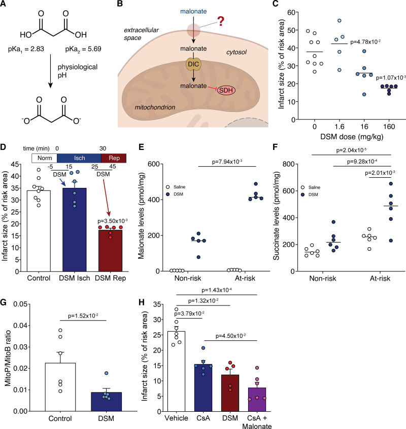
Background: Inhibiting SDH (succinate dehydrogenase), with the competitive inhibitor malonate, has shown promise in ameliorating ischemia/reperfusion injury. However, key for translation to the clinic is understanding the mechanism of malonate entry into cells to enable inhibition of SDH, its mitochondrial target, as malonate itself poorly permeates cellular membranes. The possibility of malonate selectively entering the at-risk heart tissue on reperfusion, however, remains unexplored.

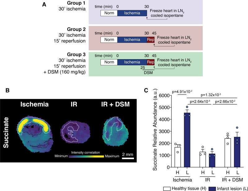
Methods: C57BL/6J mice, C2C12 and H9c2 myoblasts, and HeLa cells were used to elucidate the mechanism of selective malonate uptake into the ischemic heart upon reperfusion. Cells were treated with malonate while varying pH or together with transport inhibitors. Mouse hearts were either perfused ex vivo (Langendorff) or subjected to in vivo left anterior descending coronary artery ligation as models of ischemia/reperfusion injury. Succinate and malonate levels were assessed by liquid chromatography-tandem mass spectrometry LC-MS/MS, in vivo by mass spectrometry imaging, and infarct size by TTC (2,3,5-triphenyl-2H-tetrazolium chloride) staining.

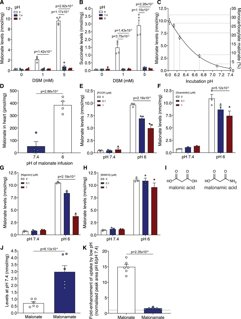
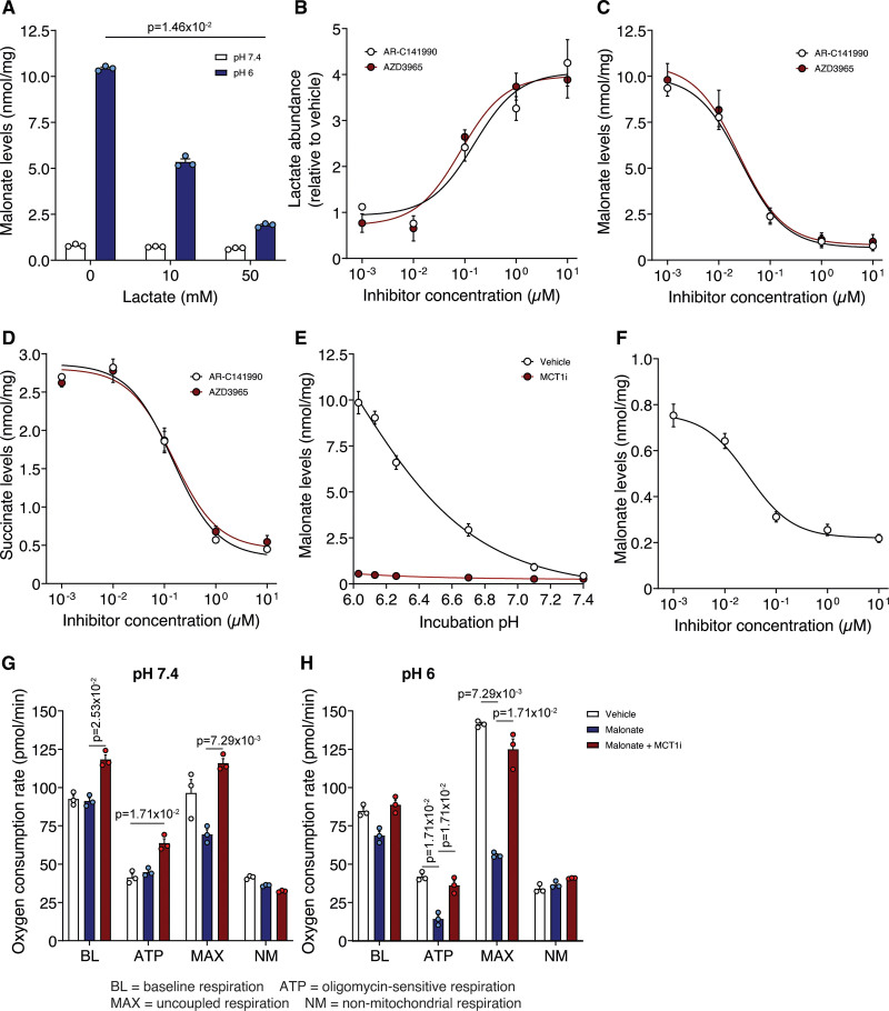
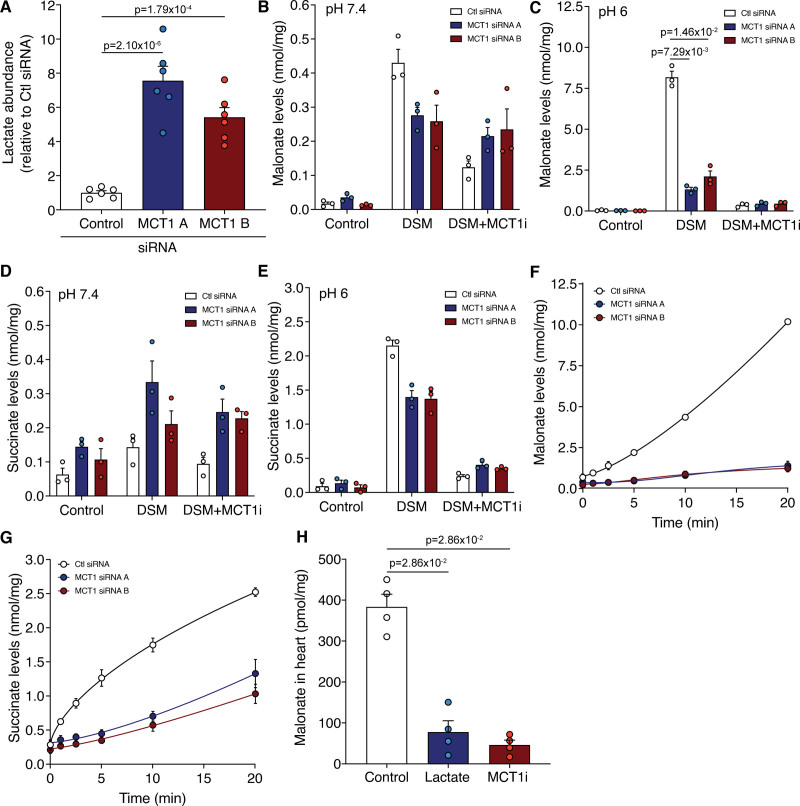
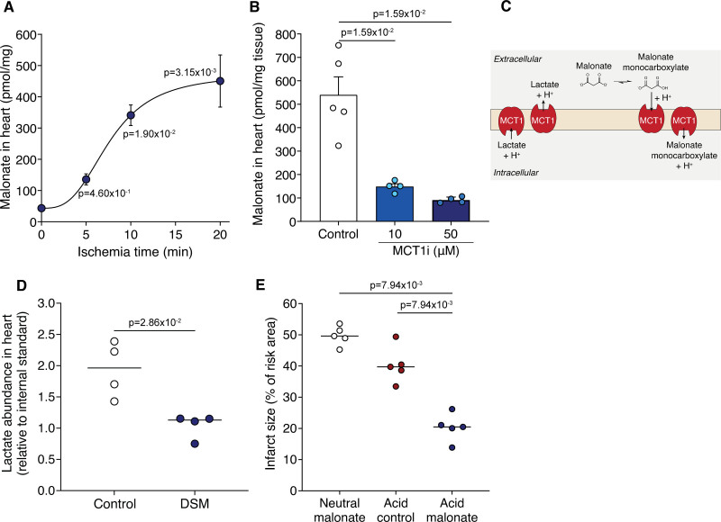
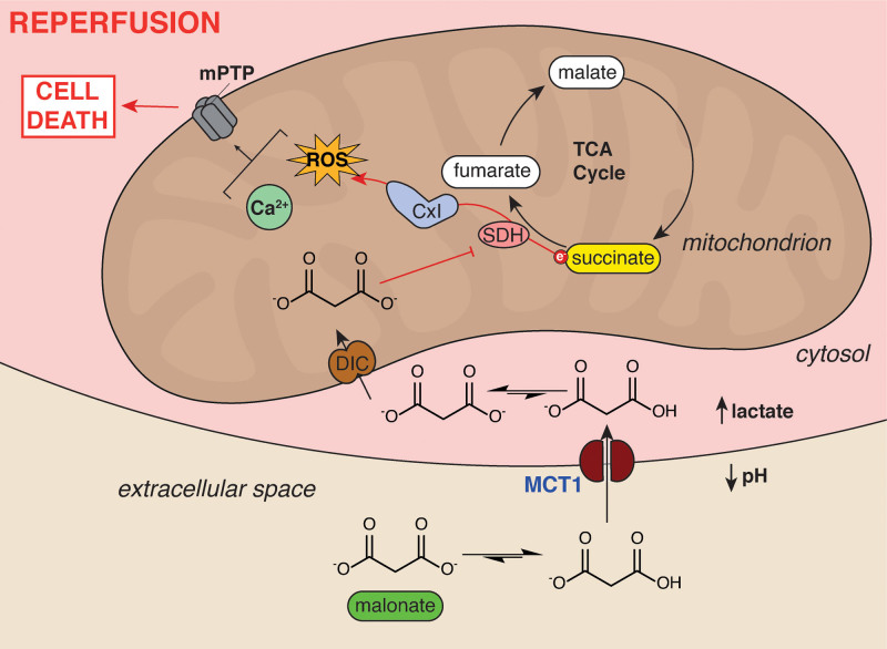
Results: Malonate was robustly protective against cardiac ischemia/reperfusion injury, but only if administered at reperfusion and not when infused before ischemia. The extent of malonate uptake into the heart was proportional to the duration of ischemia. Malonate entry into cardiomyocytes in vivo and in vitro was dramatically increased at the low pH (≈6.5) associated with ischemia. This increased uptake of malonate was blocked by selective inhibition of MCT1 (monocarboxylate transporter 1). Reperfusion of the ischemic heart region with malonate led to selective SDH inhibition in the at-risk region. Acid-formulation greatly enhances the cardioprotective potency of malonate.

Conclusions: Cardioprotection by malonate is dependent on its entry into cardiomyocytes. This is facilitated by the local decrease in pH that occurs during ischemia, leading to its selective uptake upon reperfusion into the at-risk tissue, via MCT1. Thus, malonate's preferential uptake in reperfused tissue means it is an at-risk tissue-selective drug that protects against cardiac ischemia/reperfusion injury.

Keywords: ischemia; mitochondria; myocardial infarction; reactive oxygen species; reperfusion.


Figures

Figure 1.
Figure 1.
Malonate is cardioprotective only when given at reperfusion. A, Malonate predominantly carries 2 negatively charged dicarboxylates at physiological pH. B, Schematic of the barriers to effective malonate delivery. C, Infarct size in murine LAD (left anterior descending coronary artery) ligation model with infusion of varying doses of disodium malonate (DSM; 0, 1.6, 16 or 160 mg/kg; n=9, 5, 6, 6 respectively) infused at reperfusion after 30 min ischemia, quantified by TTC (2,3,5-triphenyl-2H-tetrazolium chloride) staining. D, Infarct size in murine LAD model with infusion of DSM (160 mg/kg) at ischemia or at reperfusion (mean±SEM, n=8 (Control), 6 (DSM isch and DSM rep) biological replicates. E and F, Levels of malonate (E) and succinate (F) in nonrisk and at-risk tissue after 1 min reperfusion with either saline or DSM (160 mg/kg) after 30 min ischemia (mean±SEM, n=5 [E], 6 [F] biological replicates). Statistics: Kruskal-Wallis with Dunn post hoc test (C–E), 2-way ANOVA with Tukey post hoc test (F). G, MitoP/B ratio in at-risk heart tissue from LAD model after 30 min ischemia and 15 min reperfusion with either saline (Ctl) or DSM (160 mg/kg) infusion (mean±SEM, n=6 biological replicates, statistics: unpaired, 2-tailed Mann-Whitney U test) H, Infarct size in murine LAD model with infusion of vehicle (ethanol/Cremphor EL in saline)±CsA (cyclosporin A; 10 mg/kg) or DSM (160 mg/kg) at reperfusion (mean±SEM, n=7 [vehicle], 6 [CsA, CsA+malonate], 5 [DSM] biological replicates, statistics: Kruskal-Wallis with Dunn post hoc test). DIC indicates mitochondrial dicarboxylate carrier (SLC25A10); and SDH, succinate dehydrogenase.
Figure 2.
Figure 2.
Localization of succinate accumulation in heart tissue identified by mass spectrometry imaging. A, Outline of experimental groups for mass spectrometry imaging. B, Representative images of succinate abundance detected by mass spectrometry imaging in myocardial sections. Black outer line indicates the edge of the tissue slice, white inner line indicates infarct region. C, Quantification of succinate (C) identified by MSI (mean±SEM, n=3 biological replicates, statistics: Friedman paired test for healthy vs lesion and Kruskal-Wallis with Dunn post hoc test between conditions). au indicates arbitrary units; DSM, disodium malonate; H, healthy tissue; IR, ischemia/reperfusion; L, infarct lesion; and rep, reperfusion.
Figure 3.
Figure 3.
Malonate uptake is enhanced at low pH. A and B, C2C12 cells were incubated with disodium malonate (DSM; 0, 1, or 5 mmol/L) for 15 min at either pH 6, 7.4, or 8 before measuring intracellular malonate (A) and succinate (B) by LC-MS/MS (liquid chromatography-tandem mass spectrometry) (mean±SEM, n=4 biological replicates, statistics: Kruskal-Wallis with Dunn post hoc test). C, Malonate levels in C2C12 cells after incubation with DSM (5 mmol/L) for 15 min at various (patho)physiological pH (mean±SEM, n=3 biological replicates). D, Malonate levels in murine isolated Langendorff-perfused hearts treated with 5 mmol/L DSM infused at either pH 7.4 or 6 for 5 min (mean±SEM, n=4 biological replicates, statistics: unpaired, 2-tailed Mann-Whitney U test). E to H, C2C12 cells were incubated with DSM (5 mmol/L) for 15 min at either pH 6 or 7.4 in the presence of FCCP (carbonyl cyanide-p-trifluoromethoxyphenylhydrazone) (E), gramicidin (F), nigericin (G), or BAM15 (N5,N6-bis(2-Fluorophenyl)-[1,2,5]oxadiazolo[3,4-b]pyrazine-5,6-diamine) (H) before measuring intracellular malonate by LC-MS/MS (mean±SEM, n=3 biological replicates, statistics: Kruskal-Wallis with Dunn post hoc test). I, Structures of malonic acid and malonamic acid. J, Quantification of levels of malonate and malonamate in C2C12 cells after incubation (5 mmol/L DSM or disodium malonamate; 15 min) at pH 7.4 (n=6 biological replicates). K, C2C12 cells were incubated with either DSM or disodium malonamate (both 5 mmol/L) at pH 6 or 7.4 for 15 min and the intracellular levels measured by LC-MS/MS. (mean±SEM of the fold-enhancement of uptake at pH 6 vs pH 7.4, n=6 biological replicates). Statistics for J and K: unpaired, 2-tailed Student t test).
Figure 4.
Figure 4.
Inhibition of MCT1 (monocarboxylate transporter 1) prevents the enhanced uptake of malonate at lowered pH. A, Malonate uptake (5 mmol/L disodium malonate [DSM], 15 min) in C2C12 cells in the presence of lactate (0, 10 or 50 mmol/L). B, Lactate levels in C2C12 cells after 15 min treatment with varying concentrations of MCT1 inhibitors. C and D, Effect of MCT1 inhibition by AR-C141990 or AZD3965 on malonate (5 mmol/L DSM, 15 min) uptake (C) at pH 6 and subsequent succinate levels (D). E, C2C12 cells were incubated with DSM (5 mmol/L) for 15 min at various pH±10 µmol/L AR-C141990. F, MCT1 inhibition by AR-C141990 on malonate (5 mmol/L DSM, 15 min) uptake at pH 7.4 (A to F, mean±SEM, n=3 biological replicates, statistics: (A) Kruskal-Wallis with Dunn post hoc test). G and H, Effect of malonate (5 mmol/L DSM) on cellular oxygen consumption at pH 7.4 (G) or 6 (H) ±MCT1 inhibitor (10 µmol/L AR-C141990; data presented as nonmitochondrial respiration normalized mean oxygen consumption rate (OCR)±SEM of 3 biological replicates (n=12–16 technical replicates per biological replicate), statistics: Kruskal-Wallis with Dunn post hoc test). ATP indicates OCR in the presence of 1.5 µmol/L oligomycin; BL, baseline OCR; MAX, OCR in the presence of 1 µmol/L FCCP (carbonyl cyanide-p-trifluoromethoxyphenylhydrazone); and NM, OCR in the presence of 4 µg/mL rotenone and 10 µmol/L antimycin A.
Figure 5.
Figure 5.
Genetic knockdown of MCT1 (monocarboxylate transporter 1) prevents the uptake of malonate. A, Relative lactate levels in C2C12 cells treated with control or MCT1 siRNA (mean±SEM of lactate levels relative to control, n=6 biological replicates, statistics: 1-way ANOVA with Bonferroni post hoc test). B to E, Incubation of malonate (5 mmol/L disodium malonate [DSM]) in MCT1 KD cells at pH 7.4 (B and D) or 6 (C and E) for 15 min±MCT1i (10 µmol/L, MCT1 inhibitor AR-C141990). Levels of malonate (B and C) and succinate (D and E) quantified by LC-MS/MS (liquid chromatography-tandem mass spectrometry) (mean±SEM, n=3 biological replicates, statistics: Kruskal-Wallis with Dunn post hoc test). F and G, Time course of malonate uptake (5 mmol/L DSM) at pH 6 in MCT1 KD cells (F) and corresponding succinate levels (G; mean±SEM, n=3 biological replicates). H, Malonate levels in murine Langendorff hearts perfused at pH 6 for 5 min with 5 mmol/L DSM±lactate (50 mmol/L) or MCT1 inhibitor (10 µmol/L AR-C141990; MCT1i; mean±SEM, n=4, statistics: unpaired, 2-tailed Mann-Whitney U test vs control).
Figure 6.
Figure 6.
Ischemia drives malonate protonation, uptake, and cardioprotection. A, Langendorff-perfused murine hearts were held ischemic for either 0, 5, 10, or 20 min and reperfused with 5 mmol/L disodium malonate (DSM; pH 7.4) before malonate levels measured in the heart by LC-MS/MS (liquid chromatography-tandem mass spectrometry) (mean±SEM, n=4 (control, 5 min) or 6 (10 and 20 min) biological replicates, statistics: Kruskal-Wallis with Dunn post hoc test). B, Malonate levels in murine Langendorff hearts exposed to 20 min ischemia and reperfused with 5 mmol/L malonate (pH 7.4)±MCT1 (monocarboxylate transporter 1) inhibitor (10 or 50 µmol/L AR-C141990; mean±SEM, n=4–5 biological replicates, statistical significance assessed by unpaired, 2-tailed Mann-Whitney U test vs control). C, Model of potential lactate and malonate exchange during reperfusion. D, Lactate levels in the Langendorff heart after 20 min ischemia and 1 min reperfusion±5 mmol/L DSM (mean, n=4 biological replicates, statistics: 2-tailed, unpaired Mann-Whitney U test). E, Infarct size in murine LAD (left anterior descending coronary artery) ligation MI model with 100 µl bolus of 8 mg/kg DSM, pH 4 acid control or 8 mg/kg pH 4 formulated malonate at reperfusion after 30 min ischemia (mean, n=5 biological replicates, statistics: 2-tailed, unpaired Mann-Whitney U test vs acid malonate).
Figure 7.
Figure 7.
Schematic of ischemia-dependent malonate uptake via MCT1 (monocarboxylate transporter 1). The accumulation of lactate and protons in ischemic tissue and equilibration with the extracellular space facilitates protonation of malonate to its monocarboxylate form. This enables it to be an MCT1 substrate and enter cardiomyocytes upon reperfusion. Here, the malonate is transported into mitochondria by the mitochondrial dicarboxylate carrier where it can subsequently go on to inhibit SDH (succinate dehydrogenase). SDH inhibition reduces succinate oxidation, reactive oxygen species (ROS) production by reverse electron transport (RET) through complex I and opening of the mitochondrial permeability transition pore, thereby reducing cell death from ischemia/reperfusion (IR) injury. CxI indicates complex I; DIC, mitochondrial dicarboxylate carrier (SLC25A10); mPTP, mitochondrial permeability transition pore; and TCA, tricarboxylic acid.

Comment in

References

    1. Davidson SM, Ferdinandy P, Andreadou I, Bøtker HE, Heusch G, Ibáñez B, Ovize M, Schulz R, Yellon DM, Hausenloy DJ, et al. ; CARDIOPROTECTION COST Action (CA16225). Multitarget strategies to reduce myocardial ischemia/reperfusion injury: JACC Review Topic of the Week. J Am Coll Cardiol. 2019;73:89–99. doi: 10.1016/j.jacc.2018.09.086 - PubMed
    1. Yellon DM, Hausenloy DJ. Myocardial reperfusion injury. N Engl J Med. 2007;357:1121–1135. doi: 10.1056/NEJMra071667 - PubMed
    1. Murphy E, Steenbergen C. Mechanisms underlying acute protection from cardiac ischemia-reperfusion injury. Physiol Rev. 2008;88:581–609. doi: 10.1152/physrev.00024.2007 - PMC - PubMed
    1. Lesnefsky EJ, Chen Q, Tandler B, Hoppel CL. Mitochondrial dysfunction and myocardial ischemia-reperfusion: implications for novel therapies. Annu Rev Pharmacol Toxicol. 2017;57:535–565. doi: 10.1146/annurev-pharmtox-010715-103335 - PMC - PubMed
    1. Cung TT, Morel O, Cayla G, Rioufol G, Garcia-Dorado D, Angoulvant D, Bonnefoy-Cudraz E, Guérin P, Elbaz M, Delarche N, et al. . Cyclosporine before PCI in patients with acute myocardial infarction. N Engl J Med. 2015;373:1021–1031. doi: 10.1056/NEJMoa1505489 - PubMed

Publication types