. 2022 Aug 31;50(4):1143-1155.
doi: 10.1042/BST20220229.

Two-pore channels: going with the flows

Affiliations

Two-pore channels: going with the flows

Anthony J Morgan et al. Biochem Soc Trans. .

Abstract

In recent years, our understanding of the structure, mechanisms and functions of the endo-lysosomal TPC (two-pore channel) family have grown apace. Gated by the second messengers, NAADP and PI(3,5)P2, TPCs are an integral part of fundamental signal-transduction pathways, but their array and plasticity of cation conductances (Na+, Ca2+, H+) allow them to variously signal electrically, osmotically or chemically. Their relative tissue- and organelle-selective distribution, together with agonist-selective ion permeabilities provides a rich palette from which extracellular stimuli can choose. TPCs are emerging as mediators of immunity, cancer, metabolism, viral infectivity and neurodegeneration as this short review attests.

Keywords: Ca2+; NAADP; TPC; lysosomes.


Conflict of interest statement

The authors declare that there are no competing interests associated with the manuscript.

Figures
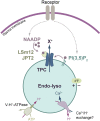
Figure 1.
Figure 1.. Second-messenger synthesis couples plasmalemmal receptors to endo-lysosomal TPCs.
A model whereby cell-surface receptors can promote the synthesis of either second messenger, the dinucleotide, NAADP or lysosome-specific lipid, PI(3,5)P2. Cations (X+) exit the acidic vesicle when TPCs are gated by NAADP via small accessory proteins, LSm12 or JPT2, or by PI(3,5)P2 that binds directly to TPCs. The lysosomal lumen is acidic by virtue of the V-H+-ATPase, which is also the primary drive of the luminally positive membrane potential (ΔΨ). In one model, the H+ gradient drives Ca2+ uptake via an unknown exchanger.
Figure 2.
Figure 2.. Distribution of TPC channels throughout the endo-lysosomal system.
TPC1 channels are predominantly found in less acidic, earlier compartments, whereas TPC2 is found in later, more acidic vesicles. Trafficking of cargo through the endo-lysosomal system (e.g. endocytosis, viruses, bacterial toxins) as well as vesicle movement and fusion is also subject to TPC control.
Figure 3.
Figure 3.. Structure of TPCs.
(A) Topology cartoon of a TPC monomer with tandem repeats of two ‘Shaker’ domains (six transmembrane domains each). The positively charged amino acids (+++) in the S4 domains confer voltage sensitivity in TPC1. Magenta branches depict luminal glycosylation. (B) Cryo-EM structure of the human TPC2 dimer (PDB: 6NQ0) with the surface structure of A and B chains in blue and pink, respectively. The lipid, PI(3,5)P2, is shown as a space-filling model (yellow, red and orange) bound to a pocket in the A chain (a second lipid molecule, bound to the equivalent pocket on the B chain, cannot be seen behind). X+ represents the direction of cation flow. (C) human TPC2 as a ribbon diagram flipped 90° compared with (B) and viewed end-on from the cytosolic face. The central pore (green shading) is contributed to by both monomers. Both PI(3,5)P2 molecules bound are visible. Structures were generated using UCSF Chimera X [132].
Figure 4.
Figure 4.. Ionic permeabilities are a function of the TPC isoform and stimulus.
Models depicting the relative permeabilities to Ca2+, Na+ and H+ are conveyed by the size of the coloured plumes. For TPC2, soluble NAADP evokes TPC2 currents with comparable Ca2+ and Na+ conductances, whereas the lipid PI(3,5)P2 stimulates Na+-selective currents.
Figure 5.
Figure 5.. Models of different ionic signalling modalities for TPC2.
With NAADP as the messenger, it binds to its accessory protein (LSm12 or JPT2, red hexagons) to evoke local Ca2+ nanodomains that are uniquely sensed by closely associated Ca2+-binding proteins (‘decoders’, brown hexagon)). When PI(3,5)P2 is the stimulus, Na+-selective currents are evoked which can depolarise the lysosome (ΔΨ) or promote osmotic changes and vesicle shrinkage (by Cl co-transport and concomitant water loss). NAADP can also promote H+ efflux through TPC2 and increase the lysosomal luminal pH (pHL).

References

    1. Morgan, A.J., Davis, L.C., Ruas, M. and Galione, A. (2015) TPC: the NAADP discovery channel? Biochem. Soc. Trans. 43, 384–389 10.1042/BST20140300 - DOI - PubMed
    1. Morgan, A.J., Platt, F.M., Lloyd-Evans, E. and Galione, A. (2011) Molecular mechanisms of endolysosomal Ca2+ signalling in health and disease. Biochem. J. 439, 349–374 10.1042/BJ20110949 - DOI - PubMed
    1. Narayanaswamy, N., Chakraborty, K., Saminathan, A., Zeichner, E., Leung, K., Devany, J.et al. (2019) A pH-correctable, DNA-based fluorescent reporter for organellar calcium. Nat. Methods 16, 95–102 10.1038/s41592-018-0232-7 - DOI - PMC - PubMed
    1. Melchionda, M., Pittman, J.K., Mayor, R. and Patel, S. (2016) Ca2+/H+ exchange by acidic organelles regulates cell migration in vivo. J. Cell Biol. 212, 803–813 10.1083/jcb.201510019 - DOI - PMC - PubMed
    1. Garrity, A.G., Wang, W., Collier, C.M., Levey, S.A., Gao, Q. and Xu, H. (2016) The endoplasmic reticulum, not the pH gradient, drives calcium refilling of lysosomes. eLife 5, e15887 10.7554/eLife.15887 - DOI - PMC - PubMed