. 2022 Aug 12;8(32):eabo7737.
doi: 10.1126/sciadv.abo7737. Epub 2022 Aug 12.

The wound-activated ERF15 transcription factor drives Marchantia polymorpha regeneration by activating an oxylipin biosynthesis feedback loop

Affiliations

The wound-activated ERF15 transcription factor drives Marchantia polymorpha regeneration by activating an oxylipin biosynthesis feedback loop

Yuanke Liang et al. Sci Adv. .

Abstract

The regenerative potential in response to wounding varies widely among species. Within the plant lineage, the liverwort Marchantia polymorpha displays an extraordinary regeneration capacity. However, its molecular pathways controlling the initial regeneration response are unknown. Here, we demonstrate that the MpERF15 transcription factor gene is instantly activated after wounding and is essential for gemmaling regeneration following tissue incision. MpERF15 operates both upstream and downstream of the MpCOI1 oxylipin receptor by controlling the expression of oxylipin biosynthesis genes. The resulting rise in the oxylipin dinor-12-oxo-phytodienoic acid (dn-OPDA) levels results in an increase in gemma cell number and apical notch organogenesis, generating highly disorganized and compact thalli. Our data pinpoint MpERF15 as a key factor activating an oxylipin biosynthesis amplification loop after wounding, which eventually results in reactivation of cell division and regeneration. We suggest that the genetic networks controlling oxylipin biosynthesis in response to wounding might have been reshuffled over evolution.


Figures
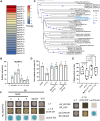
Fig. 1.
Fig. 1.. Marchantia MpERF15 shows functional conservation with Arabidopsis ERF109.
(A) Heatmap of MpERF expression after wounding. Fold changes (FCs) represent log2 values of the number of transcripts per kilobase of exon model per million mapped reads of 14-day-old Tak-1 thallus 1 hour after wounding compared to unwounded. (B) RT-qPCR analysis of MpERF15 transcripts in 10-day-old Tak-1 gemmalings following tissue incision (n = 3, six plants per replicate). (C) Phylogenetic analysis of Arabidopsis subfamily X and Marchantia ERFs. (D) Percentages of Col-0, erf109, ERF109pro:MpERF15erf109, and ERF109pro:ERF109erf109 that regenerated adventitious roots on leaf explants following transfer for 8 days on B5 medium (n = 3, >16 plants per replicate). Letters indicate significant differences [one-way analysis of variance (ANOVA)/post hoc Tukey honestly significant difference (HSD) test P < 0.05] between genotypes. (E) Root tip excision recovery of 7-day-old Col-0, erf109, ERF109pro:MpERF15erf109#2, and ERF109pro:ERF109erf109#2 at 72 hours after removing the 200-μm distal root tip (n = 4, >16 plants per replicate). Significance was calculated using Fisher’s exact test. (F) Yeast two-hybrid assays showing AtJAZ3, AtJAZ4, AtJAZ9, and MpJAZ interaction with MpERF15, and MpJAZ with AtERF109. Yeasts were grown onto SD/-Trp/-Leu (-L-T) and SD/-His/-Trp/-Leu (-L-T-H) with 2 mM 3AT media. Blue LacZ/X-Gal test indicates a positive interaction. Negative controls: GUS BD + MpERF15-AD and GUS-BD + AtERF109-AD. AD, GAL4 activation domain; BD, GAL4 binding domain.
Fig. 2.
Fig. 2.. MpERF15 is essential for gemmaling regeneration after wounding.
(A) Two-day-old gemmalings were excised along the red dashed lines to prepare pieces without apical notches (shadowed). (B) Time-lapse registration over 4 days of the regeneration of 2-day-old Tak-1 and Mperf15ko gemmalings excised as shown in (A) under continuous white light. Scale bar, 1 mm. (C) Quantification of Tak-1 and Mperf15ko regenerated apical notches 4 days post-cut (dpc). Regeneration was calculated by plotting the ratio of 4 dpc minus 0 dpc to 0 dpc explant sizes. Data represent means ± SD. Dots represent individual data points (n > 20). Statistical differences of each Mperf15ko mutant compared to Tak-1 were evaluated with Student’s t test; ****P < 0.0001. (D) RT-qPCR analysis of MpASA1 in Tak-1 and Mperf15ko mutants from t0 to 4 hours after wounding. Data represent the mean relative expression compared to the unwounded Tak-1 ± SD (n = 3, six plants per replicate); the asterisks indicate significant differences (one-way ANOVA/post hoc Tukey HSD test) of each Mperf15ko mutant compared to Tak-1.
Fig. 3.
Fig. 3.. MpERF15 drives expression of oxylipin biosynthesis genes.
(A) Overlap of genes down-regulated in Mperf15ko before wounding, MpCOI1-dependent genes, and genes up-regulated in Mpjaz-1. (B) RT-qPCR analysis of MpERF15 transcript levels in 14-day-old Tak-1 and Mpcoi1-2 thallus in unwounded control conditions (C) and 1 hour after wounding (W). (C) RT-qPCR analysis of MpCOI1 transcript levels in Tak-1 and Mperf15ko after wounding from t0 to 4 hours. (D) Overlap of genes up-regulated in Tak-1 1 hour after wounding and down-regulated in Mperf15ko in control conditions or 1 hour after wounding. (E and F) RT-qPCR analysis of MpAOS2 (E) and MpAOC (F) in 14-day-old Tak-1, Mpcoi1-2, and Mperf15ko#9 thallus in control conditions and 1 hour after wounding. (G and H) Normalized abundance of OPDA (G) and dn-OPDA (H) compounds in Tak-1 and Mperf15ko in control conditions and 1 hour after wounding in ESI− mode. (I) Effect of apical notch excision on the regeneration of 2-day-old Tak-1, Mpcoi1-2, and Mpaocko control (C) gemmalings compared with those treated for 2 hours with 10 nM OPDA (O), n > 15. (B and G to I) Asterisks indicate significant differences by Student’s t test. ns, nonsignificant. (C, E, F, and I) The different letters indicate significant differences (two-way ANOVA/post hoc Tukey HSD test, P < 0.05) between each genotype.
Fig. 4.
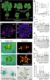
Fig. 4.. MpERF15 causes an increase in apical notches.
(A) Phenotypic comparisons of 28-day-old Tak-1, ERF109OE, and MpERF15OE plants. Scale bar, 1 cm. (B) Thallus area of individual Tak-1 and MpERF15OE lines grown for 3, 5, 7, 9, and 14 days (n > 5). Significance was calculated on the basis of two-way ANOVA followed by Tukey’s post: ***P < 0.001 and ****P < 0.0001. (C to F) Number of cells in the top epidermal cell layer (C), gemma size (D), average cell size (E), and the number of apical notches per gemma (F) of 0-day-old Tak-1, MpERF15OE, and Mperf15ko gemmae (n > 8). Significance was calculated on the basis of one-way ANOVA followed by Tukey’s post hoc test compared to Tak-1. **P < 0.01 and ****P < 0.0001. (G and H) Confocal imaging of 0-day-old gemmae with proMpEF1α:Tdtomato-N7 in a Tak-1 (G) or MpERF15OE (H) background. Scale bars, 0.1 mm. (I) Bright-field and Tdtomato (red) fluorescence images of 8-day-old gemmalings from Tak-1 and MpERF15OE. Scale bars, 0.5 mm. (G to I) The white arrowheads indicate the apical notches, and asterisks indicate the stalk cells. (J and K) proMpYUC2:GUS staining of 10-day-old thallus in a Tak-1 or MpERF15OE background. The black arrowheads indicate the blue GUS dots. Scale bars, 1 mm.
Fig. 5.
Fig. 5.. MpERF15-induced phenotypes depend on MpCOI1.
(A to D) One-day-old gemmae from Tak-1 (A), MpERF15OE#1 (B), Mpcoi1-2 (C), and MpERF15OE/Mpcoi1-2#1 (D) labeled by EdU to visualize cell cycle activity and stained with calcofluor white to visualize cell walls. Scale bars, 0.2 mm. The white arrowheads indicate EdU-positive signals, and the asterisks indicate the stalk cells. (E to H) Number of apical notches per gemma (E), number of cells in the top epidermal cell layer (F), gemma size (G), and average cell size (H) of 0-day-old Mpcoi1-2 and MpERF15OE/Mpcoi1-2 gemmae (n > 12). Significance was calculated on the basis of one-way ANOVA followed by Tukey’s post hoc test compared to Mpcoi1-2. (I) RT-qPCR analysis of MpERF15 transcripts in 14-day-old Tak-1, Mpcoi1-2, and independent MpERF15OE/Mpcoi1-2 lines. (J and K) RT-qPCR analysis of oxylipin biosynthesis–related genes MpAOS2 (J) and MpAOC (K) in 14-day-old Tak-1, MpERF15OE, Mpcoi1-2, and MpERF15OE/Mpcoi1-2. The different letters indicate significant differences (one-way ANOVA/post hoc Tukey HSD test, P < 0.01) between each genotype.
Fig. 6.
Fig. 6.. MpERF15 induces dn-OPDA biosynthesis.
(A) Normalized abundance of dn-OPDA levels in 14-day-old Tak-1 and MpERF15OE in negative (ESI−) ionization mode. Data represent means ± SD (n = 5, 12 plants per replicate). Significance was calculated on the basis of one-way ANOVA/post hoc Tukey HSD test of each genotype compared to Tak-1, *P < 0.05 and **P < 0.01. (B) Growth inhibitory effect of 10 μM OPDA/dn-OPDA on 10-day-old Marchantia gemmalings of WT Tak-1, Mpcoi1-2, Mperf15ko#9, MpERF15OE#7, and MpERF15OE/Mpcoi1-2#1. Scale bar, 1 cm. (C and D) Percentage of thalli growth inhibition of WT Tak-1, Mpcoi1-2, Mperf15ko#9, MpERF15OE#7, and MpERF15OE/Mpcoi1-2#1 by 10 μM OPDA (C) and dn-OPDA (D) compared to growth on control medium. Data represent medians (n = 3, >5 plants per replicate). The different letters indicate significant differences (one-way ANOVA/post hoc Tukey HSD test, P < 0.01) between each genotype.
Fig. 7.
Fig. 7.. MpERF15 is part of an MpCOI1-(dn-)OPDA–dependent feedback loop.
(A) RT-qPCR analysis of MpERF15 in 14-day-old WT Tak-1 thalli after transfer to control medium (EtOH) or medium supplemented with 10 μM OPDA for 2 hours. Data represent the mean relative expression compared to Tak-1 ± SD (n = 3, six plants per replicate). Statistical differences were evaluated using Student’s t test, ***P < 0.001. (B and C) RT-qPCR analysis of MpERF15 (B) and MpERF15 3′UTR. (C) Transcript levels in 7-day-old Tak-1, Mpcoi1-2, MpERF15OE#7, and MpERF15OE/Mpcoi1-2#1. Data represent the mean relative expression compared to Tak-1 ± SD (n = 3, six plants per replicate). The different letters indicate significant differences (one-way ANOVA/post hoc Tukey HSD test, P < 0.01) of each genotype. (D) Schematic diagram of MpERF15 activating a positive OPDA and dn-OPDA amplification loop that eventually results in reactivation of cell division and regeneration. Arrows represent activation; the dashed arrow points to likely control by another regulator; bar-headed lines represent repression; the line represents interactions between MpERF15 and MpJAZ; the wound-derived MpERF15-activating signal is still unknown (indicated by question mark).
Fig. 8.
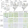
Fig. 8.. Arabidopsis ERF109 does not control jasmonate biosynthesis genes.
(A) Heatmap of RT-qPCR analysis of a subset of JA biosynthesis genes in Col-0, erf109, and ERF109pro:ERF109erf109 lines of 14-day-old unwounded and wounded seedlings (n = 3, 15 seedlings per repeat), harvested 1 hour after wounding. The FC of expression levels was normalized with log2 scale. Colors indicate up-regulated (red) and down-regulated genes (blue). (B) Five-day-old Arabidopsis Col-0, coi1-2, erf109, and ERF109pro:MpERF15erf109 seedlings grown for 7 days on control medium (−) or medium supplemented with 1, 10, or 50 μM JA. Scale bar, 1 cm. (C) Quantification of root length of seedlings shown in (B). Data represent means ± SD. Dots represent individual data points (n > 22). The different letters indicate significant differences (one-way ANOVA/post hoc Tukey HSD test, P < 0.01) of each genotype.

Similar articles

Cited by

References

    1. Marhava P., Hoermayer L., Yoshida S., Marhavý P., Benková E., Friml J., Re-activation of stem cell pathways for pattern restoration in plant wound healing. Cell 177, 957–969.e13 (2019). - PMC - PubMed
    1. Heyman J., Canher B., Bisht A., Christiaens F., De Veylder L., Emerging role of the plant ERF transcription factors in coordinating wound defense responses and repair. J. Cell Sci. 131, jcs208215 (2018). - PubMed
    1. Ikeuchi M., Favero D. S., Sakamoto Y., Iwase A., Coleman D., Rymen B., Sugimoto K., Molecular mechanisms of plant regeneration. Annu. Rev. Plant Biol. 70, 377–406 (2019). - PubMed
    1. Ikeuchi M., Ogawa Y., Iwase A., Sugimoto K., Plant regeneration: Cellular origins and molecular mechanisms. Development 143, 1442–1451 (2016). - PubMed
    1. Bannoud F., Bellini C., Adventitious rooting in Populus species: Update and perspectives. Front. Plant Sci. 12, 668837 (2021). - PMC - PubMed