

A Chemical Proteomic Map of Heme-Protein Interactions
- PMID: 35960875
- PMCID: PMC9811995
- DOI: 10.1021/jacs.2c06104
A Chemical Proteomic Map of Heme-Protein Interactions
Abstract
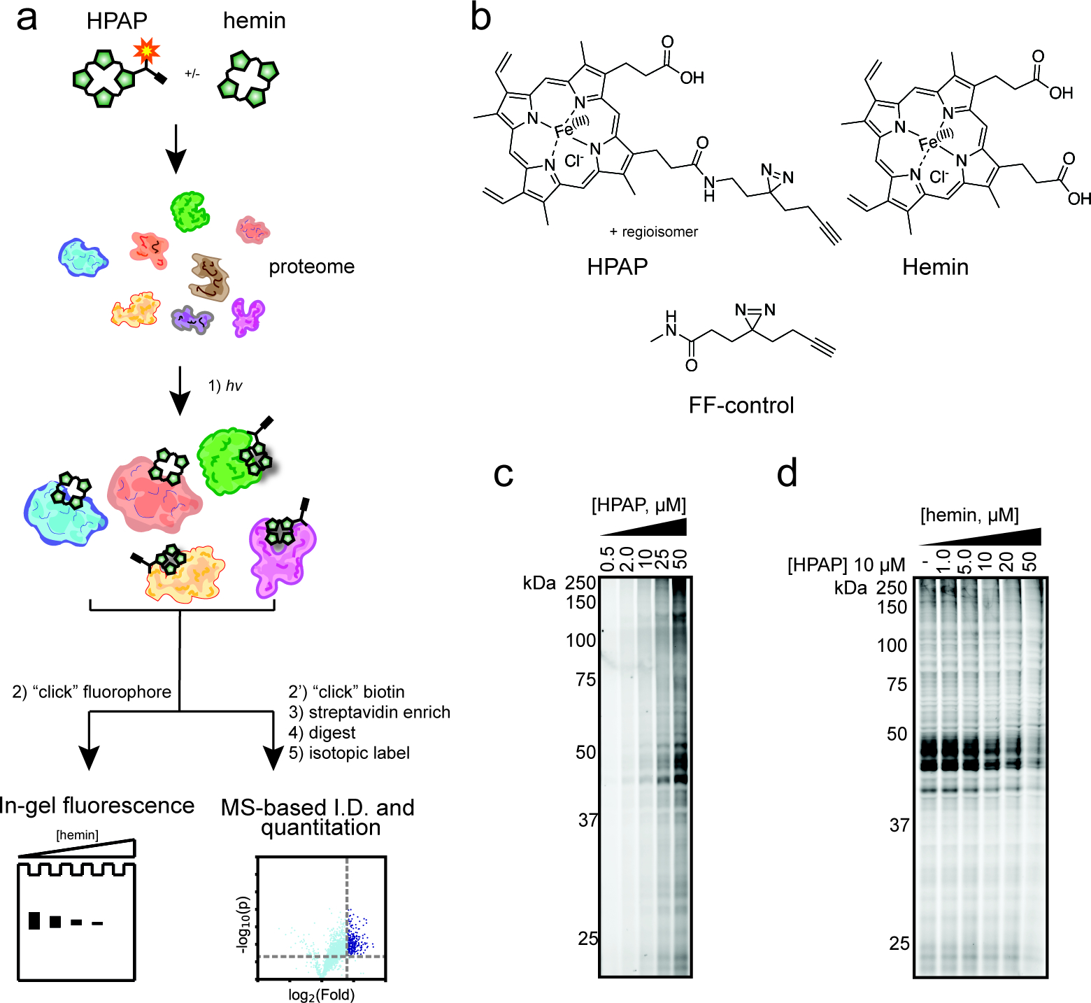
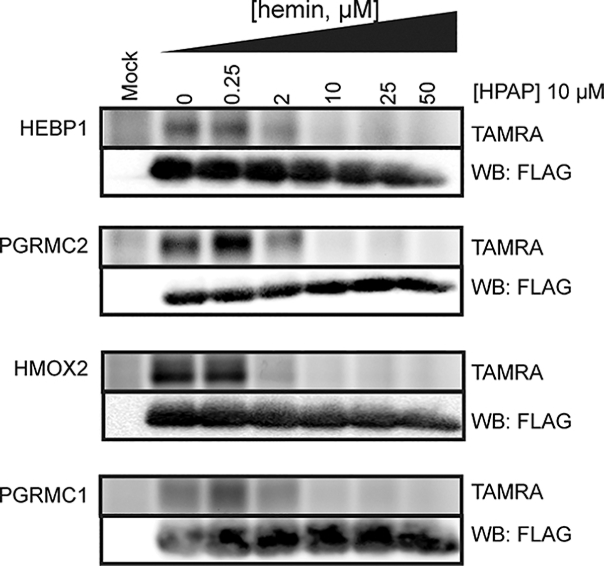
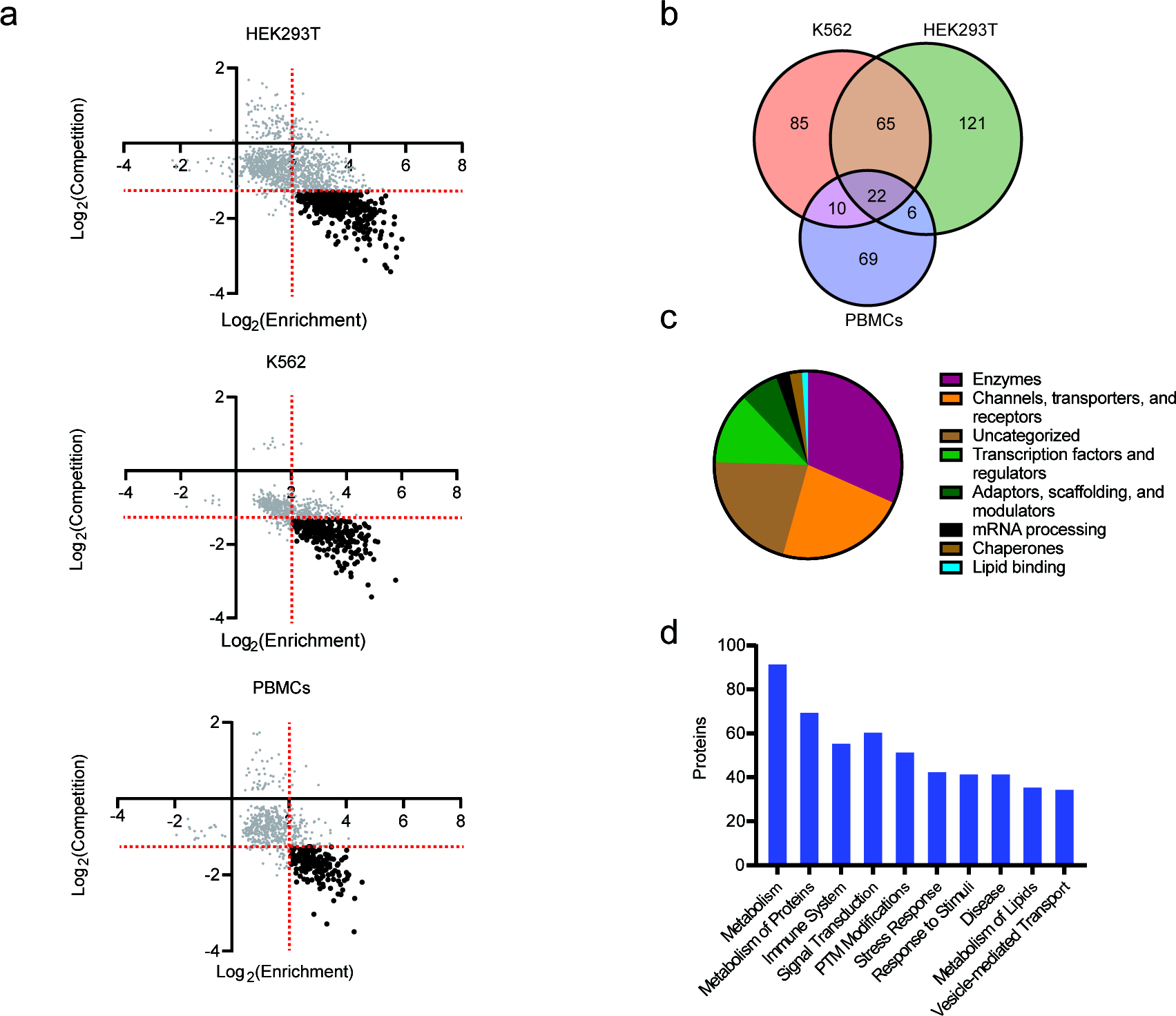
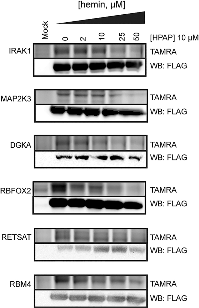
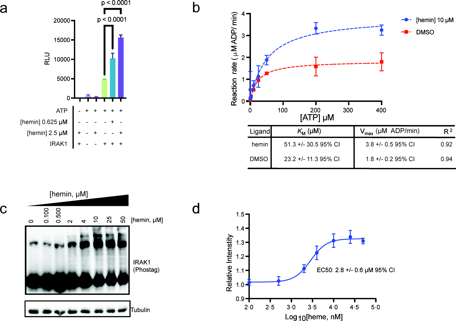
Heme is an essential cofactor for many human proteins as well as the primary transporter of oxygen in blood. Recent studies have also established heme as a signaling molecule, imparting its effects through binding with protein partners rather than through reactivity of its metal center. However, the comprehensive annotation of such heme-binding proteins in the human proteome remains incomplete. Here, we describe a strategy which utilizes a heme-based photoaffinity probe integrated with quantitative proteomics to map heme-protein interactions across the proteome. In these studies, we identified 350+ unique heme-protein interactions, the vast majority of which were heretofore unknown and consist of targets from diverse functional classes, including transporters, receptors, enzymes, transcription factors, and chaperones. Among these proteins is the immune-related interleukin receptor-associated kinase 1 (IRAK1), where we provide preliminary evidence that heme agonizes its catalytic activity. Our findings should improve the current understanding of heme's regulation as well as its signaling functions and facilitate new insights of its roles in human disease.
Conflict of interest statement
The authors declare no competing financial interest.
Figures
Similar articles
-
Depletion assisted hemin affinity (DAsHA) proteomics reveals an expanded landscape of heme-binding proteins in the human proteome.Metallomics. 2023 Mar 6;15(3):mfad004. doi: 10.1093/mtomcs/mfad004. Metallomics. 2023. PMID: 36669767 Free PMC article.
-
Profiling the Heme-Binding Proteomes of Bacteria Using Chemical Proteomics.Angew Chem Int Ed Engl. 2023 Feb 20;62(9):e202212111. doi: 10.1002/anie.202212111. Epub 2023 Jan 23. Angew Chem Int Ed Engl. 2023. PMID: 36495310
-
Heme: emergent roles of heme in signal transduction, functional regulation and as catalytic centres.Chem Soc Rev. 2019 Dec 9;48(24):5624-5657. doi: 10.1039/c9cs00268e. Chem Soc Rev. 2019. PMID: 31748766 Review.
-
Proteome-wide mapping of cholesterol-interacting proteins in mammalian cells.Nat Methods. 2013 Mar;10(3):259-64. doi: 10.1038/nmeth.2368. Epub 2013 Feb 10. Nat Methods. 2013. PMID: 23396283 Free PMC article.
-
Heme: a versatile signaling molecule controlling the activities of diverse regulators ranging from transcription factors to MAP kinases.Cell Res. 2006 Aug;16(8):681-92. doi: 10.1038/sj.cr.7310086. Cell Res. 2006. PMID: 16894358 Review.
Cited by
-
Heme Interactions as Regulators of the Alternative Pathway Complement Responses and Implications for Heme-Associated Pathologies.Curr Issues Mol Biol. 2023 Jun 16;45(6):5198-5214. doi: 10.3390/cimb45060330. Curr Issues Mol Biol. 2023. PMID: 37367079 Free PMC article. Review.
-
Hyperoxidized Species of Heme Have a Potent Capacity to Induce Autoreactivity of Human IgG Antibodies.Int J Mol Sci. 2023 Feb 8;24(4):3416. doi: 10.3390/ijms24043416. Int J Mol Sci. 2023. PMID: 36834827 Free PMC article.
-
Shapes and Patterns of Heme-Binding Motifs in Mammalian Heme-Binding Proteins.Biomolecules. 2023 Jun 23;13(7):1031. doi: 10.3390/biom13071031. Biomolecules. 2023. PMID: 37509066 Free PMC article.
-
Enhanced mapping of small-molecule binding sites in cells.Nat Chem Biol. 2024 Jul;20(7):823-834. doi: 10.1038/s41589-023-01514-z. Epub 2024 Jan 2. Nat Chem Biol. 2024. PMID: 38167919 Free PMC article.
-
Depletion assisted hemin affinity (DAsHA) proteomics reveals an expanded landscape of heme-binding proteins in the human proteome.Metallomics. 2023 Mar 6;15(3):mfad004. doi: 10.1093/mtomcs/mfad004. Metallomics. 2023. PMID: 36669767 Free PMC article.
References
-
- Gilardi G; Di Nardo G Heme iron centers in cytochrome P450: structure and catalytic activity. Rend. Lincei. 2017, 28 (1), 159–167.
-
- Lukin JA; Ho C The Structure–Function Relationship of Hemoglobin in Solution at Atomic Resolution. Chem. Rev. 2004, 104 (3), 1219–1230. - PubMed
-
- Hou SW; Reynolds MF; Horrigan FT; Heinemann SH; Hoshi T Reversible binding of heme to proteins in cellular signal transduction. Acc. Chem. Res. 2006, 39 (12), 918–924. - PubMed
-
- Shimizu T; Lengalova A; Martinek V; Martinkova M Heme: emergent roles of heme in signal transduction, functional regulation and as catalytic centres. Chem. Soc. Rev. 2019, 48 (24), 5624–5657. - PubMed
Publication types
MeSH terms
Substances
Grants and funding
LinkOut - more resources
Full Text Sources
