. 2022 Aug 24;144(33):15252-15265.
doi: 10.1021/jacs.2c05844. Epub 2022 Aug 12.

Unraveling the Pivotal Role of Atropisomerism for Cellular Internalization

Affiliations

Unraveling the Pivotal Role of Atropisomerism for Cellular Internalization

Claire Donohoe et al. J Am Chem Soc. .

Abstract

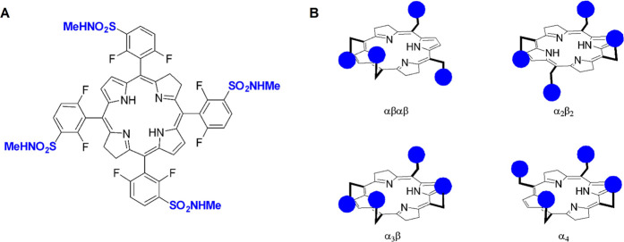
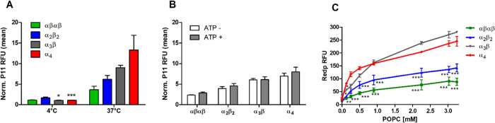
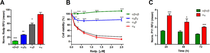
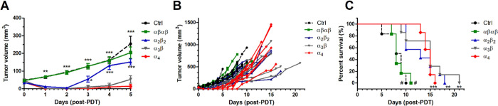
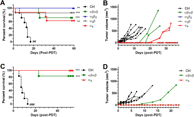
The intrinsic challenge of large molecules to cross the cell membrane and reach intracellular targets is a major obstacle for the development of new medicines. We report how rotation along a single C-C bond, between atropisomers of a drug in clinical trials, improves cell uptake and therapeutic efficacy. The atropisomers of redaporfin (a fluorinated sulfonamide bacteriochlorin photosensitizer of 1135 Da) are separable and display orders of magnitude differences in photodynamic efficacy that are directly related to their differential cellular uptake. We show that redaporfin atropisomer uptake is passive and only marginally affected by ATP depletion, plasma proteins, or formulation in micelles. The α4 atropisomer, where meso-phenyl sulfonamide substituents are on the same side of the tetrapyrrole macrocycle, exhibits the highest cellular uptake and phototoxicity. This is the most amphipathic atropisomer with a conformation that optimizes hydrogen bonding (H-bonding) with polar head groups of membrane phospholipids. Consequently, α4 binds to the phospholipids on the surface of the membrane, flips into the membrane to adopt the orientation of a surfactant, and eventually diffuses to the interior of the cell (bind-flip mechanism). We observed increased α4 internalization by cells of the tumor microenvironment in vivo and correlated this to the response of photodynamic therapy when tumor illumination was performed 24 h after α4 administration. These results show that properly orientated aryl sulfonamide groups can be incorporated into drug design as efficient cell-penetrating motifs in vivo and reveal the unexpected biological consequences of atropisomerism.


Conflict of interest statement

The authors declare the following competing financial interest(s): F.S., N.G., M.P., and L.G.A. have patents on redaporfin licensed to Luzitin SA. N.G. was an employee of Luzitin and is now an independent researcher at the University of Aveiro.

Figures

Figure 1
Figure 1
Redaporfin is a mixture of four atropisomers. (A) Redaporfin (i.e., 5,10,15,20-tetrakis(2,6-difluoro-3-N-methylsulfamoylphenyl)bacteriochlorin), molecular weight 1135 Da. (B) Atropisomers: αβαβ—two sulfonamides are on each side of the plane but in alternate positions; α2β2—two sulfonamides on each side of the plane and adjacent to each other; α3β—three sulfonamides on the same side of the plane and one on the opposite side; α4—all sulfonamides on the same side of the macrocycle plane.
Figure 2
Figure 2
Separation and X-ray crystal structures of redaporfin atropisomers. (A) Representative RP-HPLC chromatogram of redaporfin where the individual atropisomers were detected by absorption at 743 nm. (B) View of the molecular structure in the crystal of α2β2 and α4 atropisomers of P11, where the α2β2 and the α4 atropisomers were obtained from α2β2·2Me2SO and α4·4MeCN, respectively (ellipsoids are shown at the 50% probability level, and H-atoms are represented as spheres of fixed radius).
Figure 3
Figure 3
Phototoxicities of redaporfin atropisomers and of P11 atropisomers upon illumination at 740 nm (0.2 J/cm2) or 410 nm (0.0125 or 0.05 J/cm2), respectively, after 24 h of incubation. (A–C) U-2 OS, 4T1, and CT26 cells treated with redaporfin atropisomers. (D) U-2 OS cells treated with P11 atropisomers and 0.0125 J/cm2. (E–F) 4T1 and CT26 cells treated with P11 atropisomers and 0.05 J/cm2. Dose–response curves indicate the mean ± SEM of 2–3 independent experiments. Statistical significance was evaluated using two-way ANOVA vs the α4 atropisomer, * p < 0.05, ** p < 0.01, and *** p < 0.001.
Figure 4
Figure 4
Cell uptake of redaporfin and P11 atropisomers at different incubation times. (A) Fluorescence from U-2 OS cells incubated with redaporfin atropisomers (red) for 24 h, followed by nucleus staining with DAPI (blue); scale bar = 10 μm. (B–D) Cellular internalization of redaporfin atropisomers evaluated by flow cytometry at the indicated time points in U-2 OS, 4T1, and CT26 cells. (E–G) Cellular internalization of P11 atropisomers evaluated by flow cytometry at the indicated time points in U-2 OS, 4T1, and CT26 cells; bars indicate the mean ± SEM of 2–3 independent experiments; the fluorescence signal from treated cells was normalized to the untreated cells; the statistical significance was evaluated using two-way ANOVA vs the α4 atropisomer, * p < 0.05, ** p < 0.01, and *** p < 0.001.
Figure 5
Figure 5
Mechanism of atropisomer internalization in 4T1 cells. (A) P11 atropisomers (2.5 μM) incubated for 4 h at 4 °C or 37 °C or (B) incubated for 2 h upon ATP depletion; bars indicate the mean ± SEM of 2 or 3 independent experiments. (C) Fluorescence of redaporfin atropisomers (5 μM) after 20 min incubation with various concentrations of POPC liposomes; each point is mean ± SEM of 2 independent experiments; statistical significance was evaluated using two-way ANOVA vs the α4 atropisomer, * p < 0.05, ** p < 0.01, and *** p < 0.001.
Figure 6
Figure 6
Atropisomer micelle formulation cell internalization. (A) Cellular internalization of redaporfin atropisomers in vitro formulated in Kolliphor EL assessed by flow cytometry after 24 h of incubation with 4T1 cells. Bars indicate mean ± SEM of 2 independent experiments; the statistical significance was evaluated using two-way ANOVA vs the α4 atropisomer. (B) Phototoxicities of redaporfin atropisomers (formulated in Kolliphor EL) in vitro upon illumination at 740 nm (0.2 J/cm2) after 24 h of incubation with 4T1 cells; statistical significance was evaluated using two-way ANOVA vs the α4 atropisomer. (C) Level of internalization of α4 and αβαβ P11 atropisomers in single cell suspensions obtained from CT26 tumors on female BALB/c at the indicated timepoints after i.v. administration, measured by flow cytometry; bars indicate the mean ± SEM of 4–5 mice; the fluorescence signal from treated cells was normalized to the untreated cells. Significance level of the difference between the two atropisomers was evaluated via unpaired t-test, *p < 0.05, **p < 0.01, and ***p < 0.001.
Figure 7
Figure 7
Cellular-PDT (DLI = 24 h) of female BALB/c mice bearing CT26 tumors with 0.35 mg/kg redaporfin atropisomers and 60 J/cm2 at 750 nm. (A) Tumor volume represented as mean ± SEM 5 days post-PDT, which corresponds to the time interval where no mouse reached the humane endpoint; significance level vs the α4 atropisomer was evaluated by two-way ANOVA. (B) Tumor volume for each individual mouse until the humane endpoint was reached. (C) Survival curves for treatment groups with 6–7 mice. The significance level between the different treated groups was evaluated by Log-rank (Mantel–Cox) test vs Ctrl (* p < 0.05, ** p < 0.01, and *** p < 0.001) or α4 (# p < 0.001).
Figure 8
Figure 8
Vascular-PDT (DLI = 15 min) of BALB/c mice bearing CT26 tumors with 0.45 mg/kg redaporfin atropisomers and 40 J/cm2 at 750 nm. (A) Survival curves for treatment groups with 6–8 female mice. (B) Tumor volume for each individual female mouse until the endpoint was reached. (C) Survival curves for treatment groups with 6 male mice. (D) Tumor volume for each individual male mouse until the endpoint was reached. The significance level between the different treated groups was evaluated by Log-rank (Mantel–Cox) test vs Ctrl (* p < 0.05, ** p < 0.01, and *** p < 0.001) or α4 (## p < 0.01 and ### p < 0.001).

Similar articles

Cited by

References

    1. Sakamoto K.; Michibata J.; Hirai Y.; Ide A.; Ikitoh A.; Takatani-Nakase T.; Futaki S. Potentiating the Membrane Interaction of an Attenuated Cationic Amphiphilic Lytic Peptide for Intracellular Protein Delivery by Anchoring with Pyrene Moiety. Bioconjugate Chem. 2021, 32, 950–957. 10.1021/acs.bioconjchem.1c00101. - DOI - PubMed
    1. McKinlay C. J.; Waymouth R. M.; Wender P. A. Cell-Penetrating, Guanidinium-Rich Oligophosphoesters: Effective and Versatile Molecular Transporters for Drug and Probe Delivery. J. Am. Chem. Soc. 2016, 138, 3510–3517. 10.1021/jacs.5b13452. - DOI - PMC - PubMed
    1. Pei D.; Buyanova M. Overcoming Endosomal Entrapment in Drug Delivery. Bioconjate Chem. 2019, 30, 273–283. 10.1021/acs.bioconjchem.8b00778. - DOI - PMC - PubMed
    1. Guha S.; Ghimire J.; Wu E.; Wimley W. C. Mechanistic Landscape of Membrane-Permeabilizing Peptides. Chem. Rev. 2019, 119, 6040–6085. 10.1021/acs.chemrev.8b00520. - DOI - PMC - PubMed
    1. Allen J.; Najjar K.; Erazo-Oliveras A.; Kondow-McConaghy H. M.; Brock D. J.; Graham K.; Hager E. C.; Marschall A. L. J.; Dübel S.; Juliano R. L.; Pellois J.-P. Cytosolic Delivery of Macromolecules in Live Human Cells Using the Combined Endosomal Escape Activities of a Small Molecule and Cell Penetrating Peptides. ACS Chem. Biol. 2019, 14, 2641–2651. 10.1021/acschembio.9b00585. - DOI - PMC - PubMed

Publication types