

A Metabolite Produced by Gut Microbes Represses Phage Infections in Vibrio cholerae
- PMID: 35960903
- PMCID: PMC10981169
- DOI: 10.1021/acschembio.2c00422
A Metabolite Produced by Gut Microbes Represses Phage Infections in Vibrio cholerae
Abstract
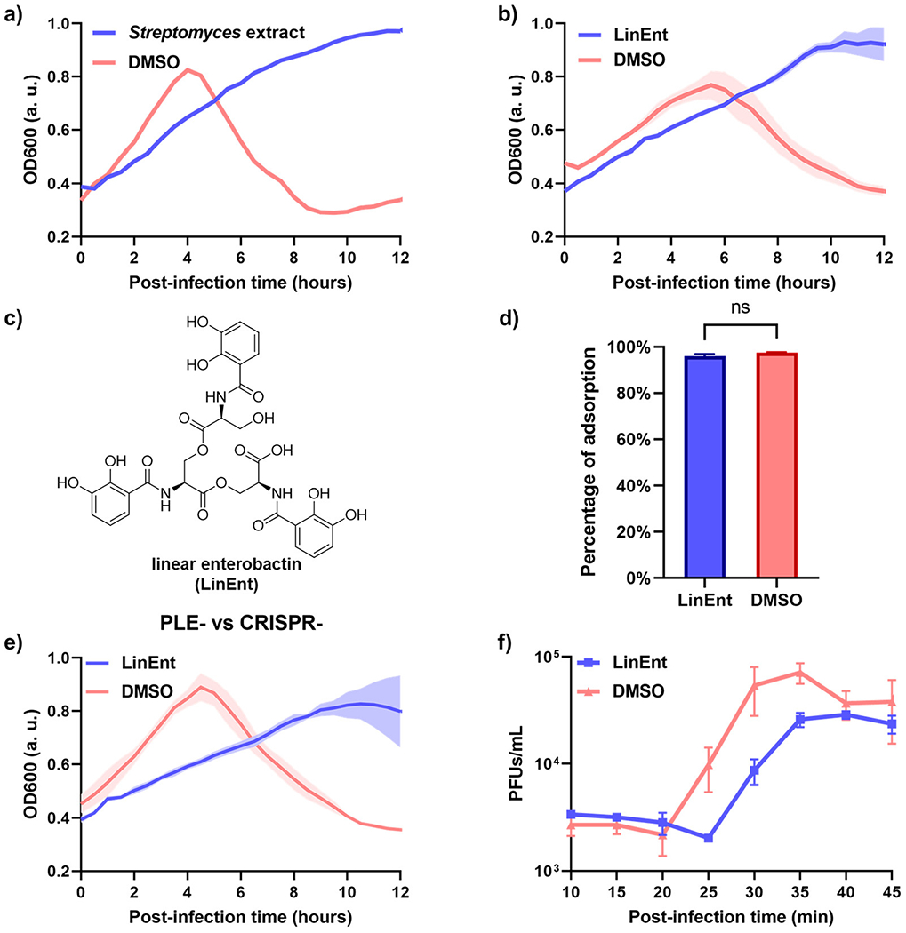
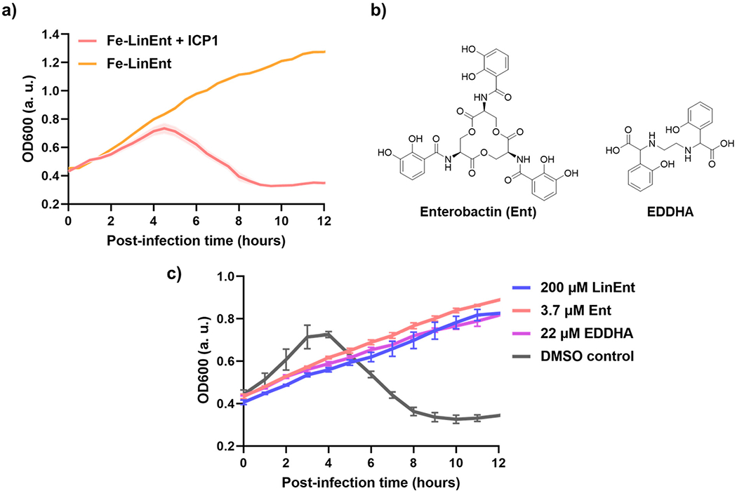
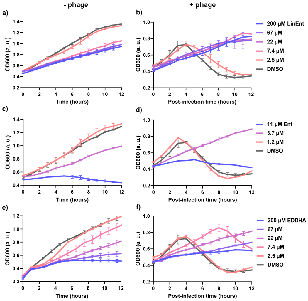
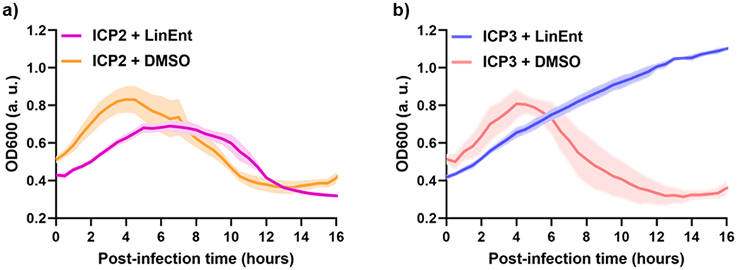
Vibrio cholerae is the causative agent of the severe diarrheal disease cholera. Bacteriophages that prey on V. cholerae may be employed as phage therapy against cholera. However, the influence of the chemical environment on the infectivity of vibriophages has been unexplored. Here, we discovered that a common metabolite produced by gut microbes─linear enterobactin (LinEnt), represses vibriophage proliferation. We found that the antiphage effect by LinEnt is due to iron sequestration and that multiple forms of iron sequestration can protect V. cholerae from phage predation. This discovery emphasizes the significance that the chemical environment can have on natural phage infectivity and phage-based interventions.
Conflict of interest statement
The authors declare no competing financial interest.
Figures
Similar articles
-
Dominant Vibrio cholerae phage exhibits lysis inhibition sensitive to disruption by a defensive phage satellite.Elife. 2020 Apr 24;9:e53200. doi: 10.7554/eLife.53200. Elife. 2020. PMID: 32329714 Free PMC article.
-
Mechanisms of the evolutionary arms race between Vibrio cholerae and Vibriophage clinical isolates.Int Microbiol. 2017 Sep;20(3):116-120. doi: 10.2436/20.1501.01.292. Int Microbiol. 2017. PMID: 29446802 Free PMC article. Review.
-
Evolutionary Sweeps of Subviral Parasites and Their Phage Host Bring Unique Parasite Variants and Disappearance of a Phage CRISPR-Cas System.mBio. 2021 Feb 22;13(1):e0308821. doi: 10.1128/mbio.03088-21. Epub 2022 Feb 15. mBio. 2021. PMID: 35164562 Free PMC article.
-
Niche adaptation limits bacteriophage predation of Vibrio cholerae in a nutrient-poor aquatic environment.Proc Natl Acad Sci U S A. 2019 Jan 29;116(5):1627-1632. doi: 10.1073/pnas.1810138116. Epub 2019 Jan 11. Proc Natl Acad Sci U S A. 2019. PMID: 30635420 Free PMC article.
-
Phage-bacterial interactions in the evolution of toxigenic Vibrio cholerae.Virulence. 2012 Nov 15;3(7):556-65. doi: 10.4161/viru.22351. Epub 2012 Oct 17. Virulence. 2012. PMID: 23076327 Free PMC article. Review.
Cited by
-
Natural products influence bacteriophage infectivity.Nat Prod Rep. 2025 Aug 18. doi: 10.1039/d5np00014a. Online ahead of print. Nat Prod Rep. 2025. PMID: 40824115 Free PMC article. Review.
-
Bacterium secretes chemical inhibitor that sensitizes competitor to bacteriophage infection.bioRxiv [Preprint]. 2024 Jan 31:2024.01.31.578241. doi: 10.1101/2024.01.31.578241. bioRxiv. 2024. Update in: Nat Microbiol. 2025 Feb;10(2):362-373. doi: 10.1038/s41564-024-01910-8. PMID: 38352521 Free PMC article. Updated. Preprint.
-
Chemical inhibition of a bacterial immune system 1.bioRxiv [Preprint]. 2025 Feb 21:2025.02.20.638879. doi: 10.1101/2025.02.20.638879. bioRxiv. 2025. PMID: 40027640 Free PMC article. Preprint.
-
Streptomyces secretes a siderophore that sensitizes competitor bacteria to phage infection.Nat Microbiol. 2025 Feb;10(2):362-373. doi: 10.1038/s41564-024-01910-8. Epub 2025 Jan 8. Nat Microbiol. 2025. PMID: 39779880
References
-
- Fleming A On the antibacterial action of cultures of a penicillium, with special reference to their use in the isolation of B. influenzæ. Br. J. Exp. Pathol 1929, 10 (3), 226–236.
-
- Summers WC Bacteriophage therapy. Annu. Rev. Microbiol 2001, 55 (1), 437–451. - PubMed
-
- Blair JMA; Webber MA; Baylay AJ; Ogbolu DO; Piddock LJV Molecular mechanisms of antibiotic resistance. Nature Reviews Microbiology 2015, 13 (1), 42–51. - PubMed
-
- Dedrick RM; Guerrero-Bustamante CA; Garlena RA; Russell DA; Ford K; Harris K; Gilmour KC; Soothill J; Jacobs-Sera D; Schooley RT; et al. Engineered bacteriophages for treatment of a patient with a disseminated drug-resistant Mycobacterium abscessus. Nature Medicine 2019, 25 (5), 730–733. - PMC - PubMed
Publication types
MeSH terms
Substances
Grants and funding
LinkOut - more resources
Full Text Sources
Medical
