. 2023 May 4;25(5):940-954.
doi: 10.1093/neuonc/noac193.

Prognostic validation of a new classification system for extent of resection in glioblastoma: A report of the RANO resect group

Affiliations

Prognostic validation of a new classification system for extent of resection in glioblastoma: A report of the RANO resect group

Philipp Karschnia et al. Neuro Oncol. .

Abstract

Background: Terminology to describe extent of resection in glioblastoma is inconsistent across clinical trials. A surgical classification system was previously proposed based upon residual contrast-enhancing (CE) tumor. We aimed to (1) explore the prognostic utility of the classification system and (2) define how much removed non-CE tumor translates into a survival benefit.

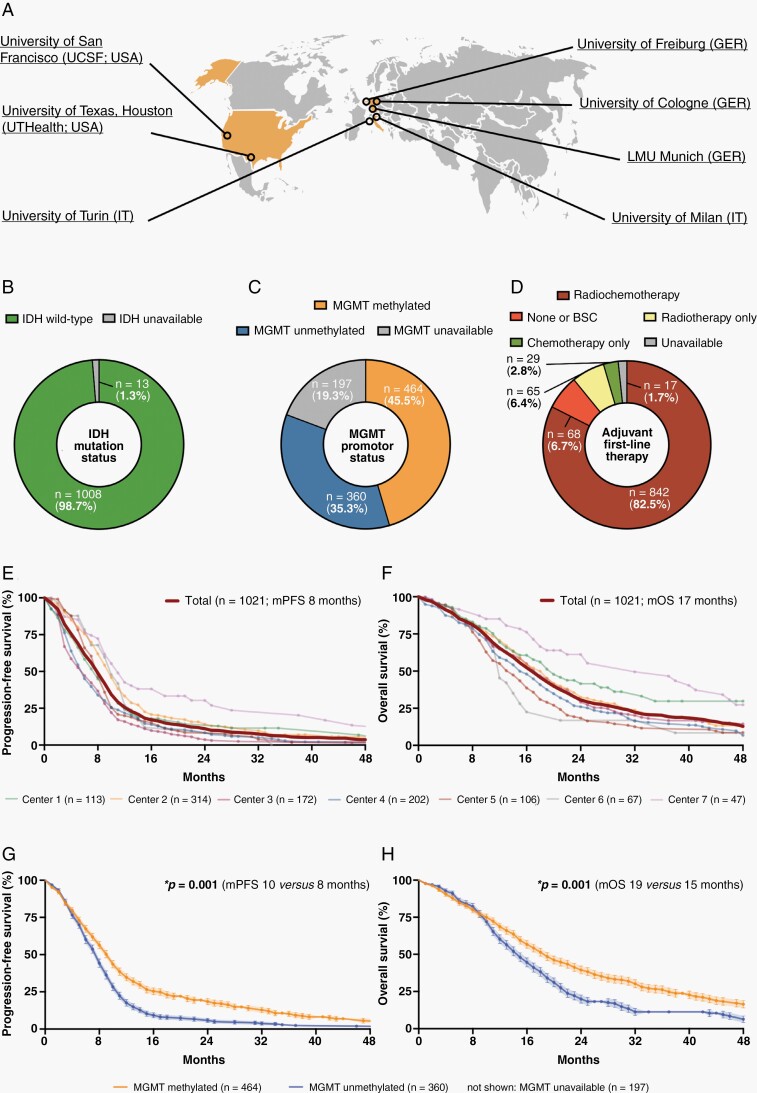
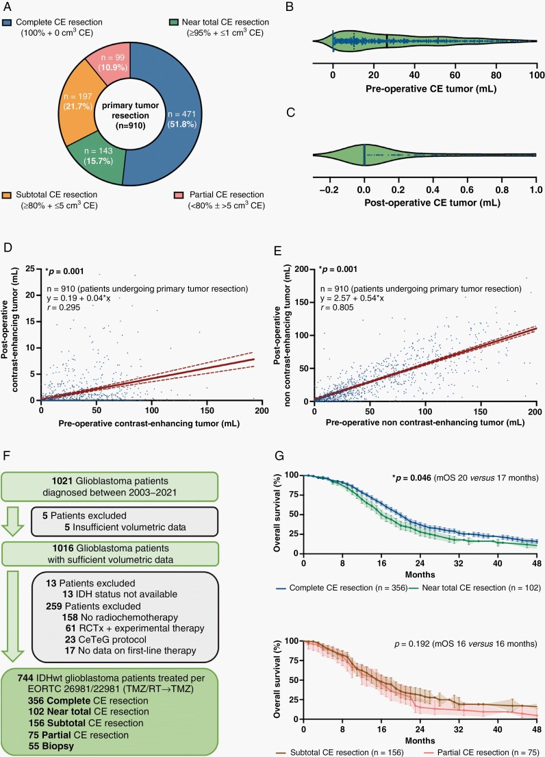
Methods: The international RANO resect group retrospectively searched previously compiled databases from 7 neuro-oncological centers in the USA and Europe for patients with newly diagnosed glioblastoma per WHO 2021 classification. Clinical and volumetric information from pre- and postoperative MRI were collected.

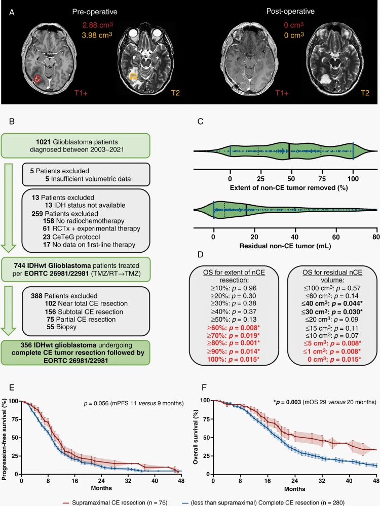
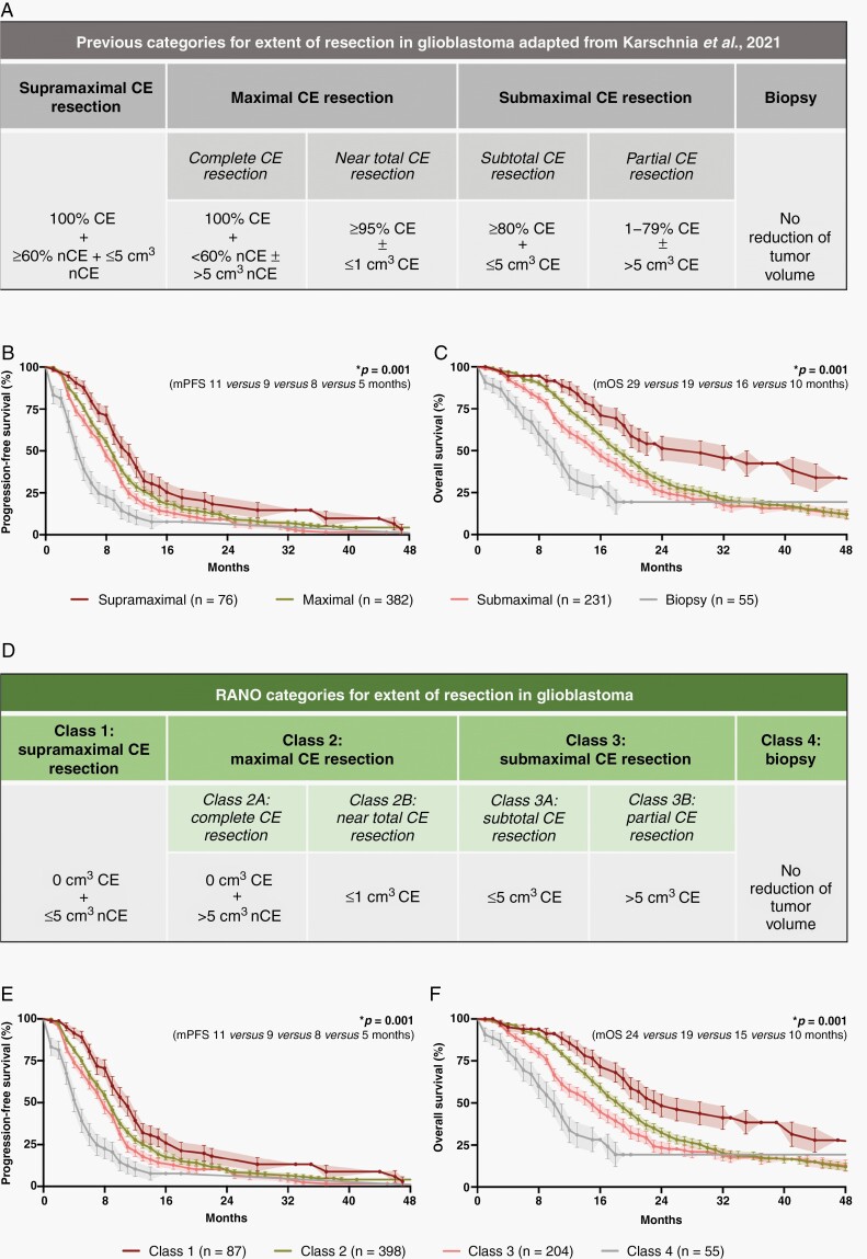
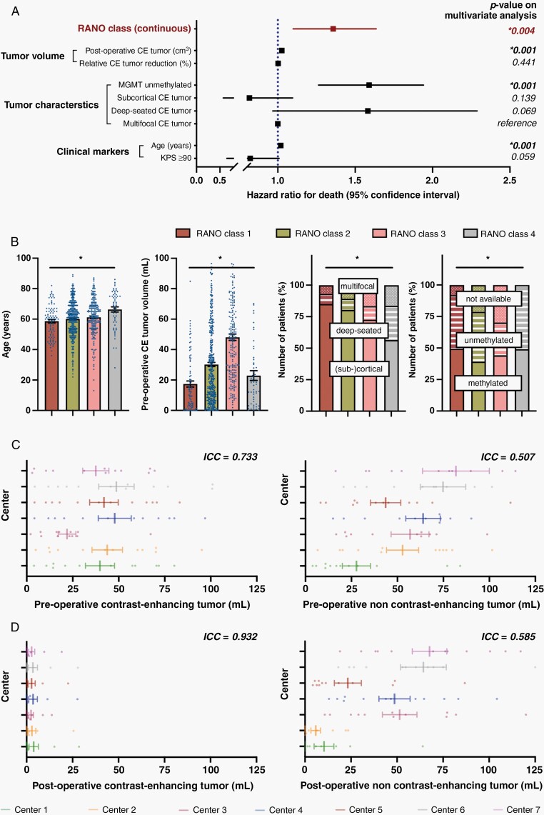
Results: We collected 1,008 patients with newly diagnosed IDHwt glioblastoma. 744 IDHwt glioblastomas were treated with radiochemotherapy per EORTC-26981/22981 (TMZ/RT→TMZ) following surgery. Among these homogenously treated patients, lower absolute residual tumor volumes (in cm3) were favorably associated with outcome: patients with "maximal CE resection" (class 2) had superior outcome compared to patients with "submaximal CE resection" (class 3) or "biopsy" (class 4). Extensive resection of non-CE tumor (≤5 cm3 residual non-CE tumor) was associated with better survival among patients with complete CE resection, thus defining class 1 ("supramaximal CE resection"). The prognostic value of the resection classes was retained on multivariate analysis when adjusting for molecular and clinical markers.

Conclusions: The proposed "RANO categories for extent of resection in glioblastoma" are highly prognostic and may serve for stratification within clinical trials. Removal of non-CE tumor beyond the CE tumor borders may translate into additional survival benefit, providing a rationale to explicitly denominate such "supramaximal CE resection."

Keywords: EOR; classification; glioblastoma; outcome; surgical resection.


Figures

Fig. 1
Fig. 1
Baseline characteristics of the entire study cohort including 1,021 glioblastoma patients. (A) Schematic localization of the 7 participating neuro-oncological centers. (B–D) Distribution of IDH mutation status (B), MGMT promotor status (C), and first-line therapies (D) across the entire study cohort (n = 1,021). (E–H) Kaplan–Meier estimates of progression-free survival (E and G) and OS (F and H) for the entire study cohort (n = 1,021). Curves are given for patients stratified according to the respective study center (E and F) and MGMT promotor status (G and H). Points: deceased/censored patients; shading: SEM.
Fig. 2
Fig. 2
Stratification according to extent of CE tumor resection and prognostic implications. (A) Stratification of all patients undergoing microsurgical tumor resection (n = 910) according to the previously proposed classification system based on extent of CE tumor resection. (B and C) Pre-(B) and postoperative CE tumor volume (C; in cm3) after microsurgical tumor resection (n = 910). Median ± 95% CI. (D and E) Simple linear regression analyses comparing the pre- to the postoperative tumor volumes for CE (D) and non-CE tumor (E) in patients undergoing microsurgical tumor resection (n = 910). Pearson correlation coefficients (r), calculated equations including slope β 1, and P-values are given. Dotted lines: 95% CI. (F) Schematic representation of the formation of a patient cohort exclusively including IDH-wildtype glioblastomas homogenously treated per EORTC-26981/22981-protocol (n = 744). (G and H) Kaplan–Meier estimates of OS in IDH-wildtype glioblastomas treated per EORTC-26981/22981-protocol (n = 744). Patients stratified to the category “complete CE resection” were compared to “near total CE resection” (G), and patients stratified to the category “subtotal CE resection” were compared to “partial CE resection” (H). Points: deceased/censored patients; shading: SEM.
Fig. 3
Fig. 3
Prognostic value of non-CE tumor resection beyond CE tumor borders. (A) Axial brain MRI with contrast-enhanced T1- (left on each panel) and T2-weighted (right on each panel) sequences demonstrating a right temporo-occipital glioblastoma. On preoperative imaging (left panel), CE (red) and non-CE tumors (orange) are delineated. Note that non-CE tumor was defined as T2/FLAIR-alterations beyond the enhancing tumor borders not corresponding to surgically induced edema or ischemia. On postoperative imaging (right panel), complete resection of CE and non-CE tumors was achieved. (B) Schematic representation of the formation of a patient cohort exclusively including IDH-wildtype glioblastomas homogenously treated per EORTC-26981/22981-protocol and in which complete CE resection was provided (n = 365). (C) Extent of non-CE tumor removed (upper panel; in percentage) and postoperative non-CE tumor volume (lower panel; in cm3) in IDH-wildtype glioblastomas treated per EORTC-26981/22981-protocol and in which complete CE resection was provided (n = 365). Median ± 95%CI. (D) Univariate analyses for OS in IDH-wildtype glioblastomas treated per EORTC-26981/22981-protocol and in which complete CE resection was provided (n = 365) when stratified according to relative reduction of non-CE tumor (left panel; in percentage) or postoperative non-CE tumor volume (right panel; in cm3). P-values are given; asterisks and bold letters indicate P ≤ .05. (E and F) Kaplan–Meier estimates of progression-free survival (E) and OS (F) in IDH-wildtype glioblastomas treated per EORTC-26981/22981-protocol and in which complete CE resection was provided (n = 365). Curves are given for patients with “supramaximal" CE resection (red; defined as ≥ 60% reduction of non-CE tumor volume + ≤5 cm3 residual non-CE tumor) and less than “supramaximal" CE resection (blue). Points: deceased/censored patients; shading: SEM.
Fig. 4
Fig. 4
Stratification systems to describe EOR in glioblastoma. (A) Previously proposed classification system by Karschnia et al. (in Eur J Cancer, 2021) based upon the relative reduction of tumor volume (in percentage) and the absolute residual tumor volume (in cm3) on postoperative MRI. (B and C): Kaplan–Meier estimates of progression-free survival (B) and OS (C) in IDH-wildtype glioblastomas treated per EORTC-26981/22981-protocol (n = 744). Patients were stratified according to the categories proposed in the classification system. Points: deceased/censored patients; shading: SEM. (D) The developed RANO categories for EOR in glioblastoma which solely base upon the absolute residual tumor volume (in cm3) on postoperative MRI. (E and F): Kaplan–Meier estimates of progression-free survival (E) and OS (F) in IDH-wildtype glioblastomas treated per EORTC-26981/22981-protocol (n = 744). Patients were stratified according to the RANO categories. Note that the prognostic value of the previously proposed classification system (A) is retained in the RANO categories (D) although the relative reduction of tumor volume (in percentage) has been removed as a classification criterion. Points: deceased/censored patients; shading: SEM.
Fig. 5
Fig. 5
Multivariate analysis and determination of inter-rater variability to control for potential confounding effects. (A) Multivariate analysis using a Cox proportional hazard regression model estimating the hazard ratio for death of numerous factors which were of significance on univariate analysis. Note that the prognostic value of the RANO classes and absolute residual CE tumor volume (in cm3) was conserved, whereas the prognostic relevance of the relative reduction of CE tumor volume (in percentage) was lost. “Multifocal CE tumor” served as reference for “deep-seated CE tumor” and “subcortical CE tumor.” Hazard ratio ± 95% CI. (B) Distribution of age (left panel), preoperative CE tumor volume (second to left panel), tumor localization (second to right panel), and MGMT promotor status (right panel) across the 4 different RANO categories. Asterisks indicate P ≤ .05 when all four groups were tested together using a Kruskal–Wallis test (for continuous data) or a χ2-test (for categorical variables). Mean ± SEM for continuous data. (C and D): Volumetric analyses of twelve identical pre-(C) and postoperative (D) MRIs by the seven participating centers (color-coded). ICCs (<0.5: poor reliability, 0.5–0.75: moderate reliability, 0.75–0–9: good reliability, >0.9: excellent reliability) are reported for pre-and postoperative CE (left panel) as well as non-CE tumor (right panel). Note that ICCs are higher for post- than for preoperative imaging and for CE than for non-CE tumor. Mean ± SEM.

Comment in

References

    1. Weller M, van den Bent M, Preusser M, et al. EANO guidelines on the diagnosis and treatment of diffuse gliomas of adulthood. Nat Rev Clin Oncol. 2020; 18(3):170–186. - PMC - PubMed
    1. Wen PY, Weller M, Lee EQ, et al. Glioblastoma in adults: a Society for Neuro-Oncology (SNO) and European Society of Neuro-Oncology (EANO) consensus review on current management and future directions. Neuro Oncol. 2020; 22(8):1073–1113. - PMC - PubMed
    1. Brown TJ, Brennan MC, Li M, et al. Association of the extent of resection with survival in glioblastoma: a systematic review and meta-analysis. JAMA Oncol. 2016; 2(11):1460–1469. - PMC - PubMed
    1. Gessler F, Bernstock JD, Braczynski A, et al. Surgery for glioblastoma in light of molecular markers: impact of resection and MGMT promoter methylation in newly diagnosed IDH-1 wild-type glioblastomas. Neurosurgery. 2019; 84(1):190–197. - PMC - PubMed
    1. Incekara F, Smits M, van der Voort SR, et al. The association between the extent of glioblastoma resection and survival in light of MGMT promoter methylation in 326 patients with newly diagnosed IDH-wildtype glioblastoma. Front Oncol. 2020; 10:1087. - PMC - PubMed