. 2022 Aug 12;5(11):e202201561.
doi: 10.26508/lsa.202201561. Print 2022 Nov.

Cdh5-lineage-independent origin of dermal lymphatics shown by temporally restricted lineage tracing

Affiliations

Cdh5-lineage-independent origin of dermal lymphatics shown by temporally restricted lineage tracing

Yan Zhang et al. Life Sci Alliance. .

Abstract

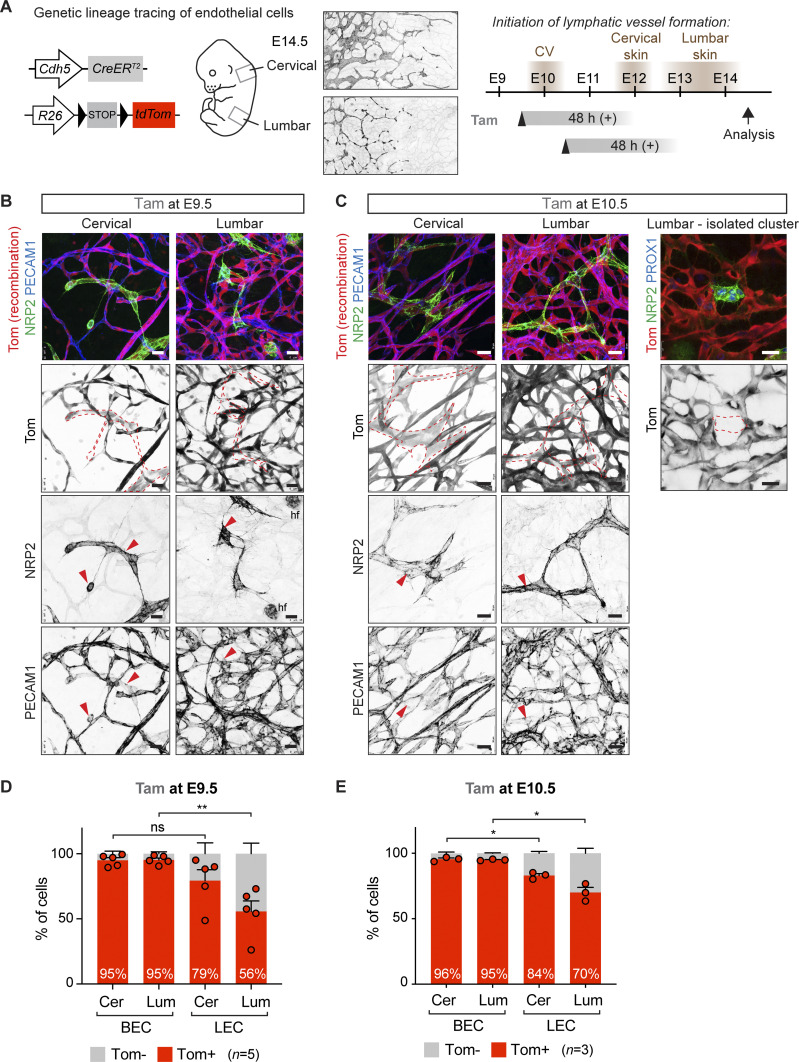
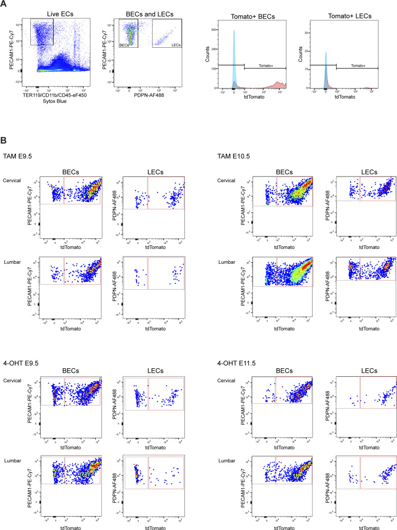
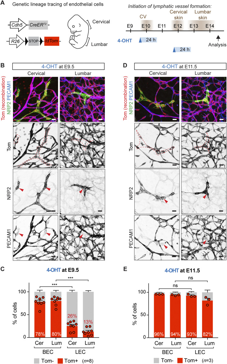
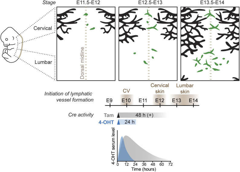
The developmental origins of lymphatic endothelial cells (LECs) have been under intense research after a century-long debate. Although previously thought to be of solely venous endothelial origin, additional sources of LECs were recently identified in multiple tissues in mice. Here, we investigated the regional differences in the origin(s) of the dermal lymphatic vasculature by lineage tracing using the pan-endothelial Cdh5-CreER T2 line. Tamoxifen-induced labeling of blood ECs at E9.5, before initiation of lymphatic development, traced most of the dermal LECs but with lower efficiency in the lumbar compared with the cervical skin. By contrast, when used at E9.5 but not at E11.5, 4-hydroxytamoxifen, the active metabolite of tamoxifen that provides a tighter window of Cre activity, revealed low labeling frequency of LECs, and lymphvasculogenic clusters in the lumbar skin in particular. Temporally restricted lineage tracing thus reveals contribution of LECs of Cdh5-lineage-independent origin to dermal lymphatic vasculature. Our results further highlight Cre induction strategy as a critical parameter in defining the temporal window for stage-specific lineage tracing during early developmental stages of rapid tissue differentiation.


Conflict of interest statement

The authors declare that they have no conflict of interest.

Figures

Figure 1.
Figure 1.. Tamoxifen-induced lineage labeling of BECs before initiation of lymphatic development.
(A) On the left: schematic of the Cdh5-CreERT2 transgene and R26-tdTom reporter for lineage tracing of BEC-derived lymphatic endothelial cells (LECs). On the right: timing of LEC differentiation from the cardinal vein (CV) and dermal lymphatic-vessel formation in the cervical and lumbar regions of the skin (shown by NRP2 whole-mount immunofluorescence), as well as the time points of tamoxifen administration and analysis are indicated. The estimated time window of Cre activity is up to at least 48 h. (B, C) Whole-mount immunofluorescence of E14.5 skin from R26-tdTom;Cdh5-CreERT2 embryos treated with tamoxifen at E9.5 (B) or at E10.5 (C). Dotted lines highlight lymphatic vessels and LEC clusters, defined by high NRP2 and low PECAM1 expression (red arrowheads). hf, hair follicle cells. (D, E) FACS analysis of ECs from R26-tdTom;Cdh5-CreERT2 embryos treated with tamoxifen at E9.5 (D) or at E10.5 (E). Note efficient Cre-mediated recombination (Tomato expression) in BECs and lower recombination in LECs in the lumbar region of skin. Cer, cervical; Lum, lumbar. (D, E) Data represent mean % of Tomato+ cells (n = 5 embryos from 3 liters (D) or n = 3 embryos from 1 liter (E), dots indicate individual embryos) ± SEM. P-value, paired two-tailed t test. *P < 0.05, **P < 0.01. Scale bars: 25 μm. Source data are available for this figure.
Figure S1.
Figure S1.. Analysis of dermal ECs by flow cytometry.
(A) Gating scheme for analysis of Tomato expression in dermal BECs (PECAM1+PDPN) and lymphatic endothelial cells (PECAM1+PDPN+) from a tamoxifen-treated R26-tdTom;Cdh5-CreERT2 embryo. (B) Representative FACS dot plots for quantifications in Figs 1D and E and 2C and E. Dot plots are displayed in the FlowJo pseudo color mode.
Figure 2.
Figure 2.. Temporally restricted lineage tracing of BEC-derived lymphatic endothelial cells (LECs) using 4-OHT.
(A) On the left: schematic of the Cdh5-CreERT2 transgene and R26-tdTom reporter for lineage tracing of BEC-derived LECs. On the right: timing of LEC differentiation from the cardinal vein (CV) and dermal lymphatic-vessel formation in the cervical and lumbar regions of the skin (depicted on the left), as well as the time points of 4-OHT administration and analysis are indicated. The estimated time window of Cre activity is up to 24 h. (B, D) Whole-mount immunofluorescence of E14.5 skin from R26-tdTom;Cdh5-CreERT2 embryos treated with 4-OHT at E9.5 (B) or at E11.5 (D). Dotted lines highlight lymphatic vessels and LEC clusters, defined by high NRP2 and low PECAM1 expression (red arrowheads). (C, E) FACS analysis of ECs from R26-tdTom;Cdh5-CreERT2 embryos treated with 4-OHT at E9.5 (C) or at E11.5 (E). Note efficient Cre-mediated recombination (Tomato expression) in BECs but low recombination in LECs in embryos treated at E9.5. Cer, cervical; Lum, lumbar. (C, E) Data represent mean % of Tomato+ cells (n = 8 embryos from 3 liters (C) or n = 3 embryos from 1 liter (E), dots indicate individual embryos) ± SEM. P-value, paired two-tailed t test. ***P < 0.001. Scale bars: 25 μm. Source data are available for this figure.
Figure 3.
Figure 3.. Stages of dermal lymphatic-vessel development in relation to the window of Cre activity induced by tamoxifen or 4-OHT to trace BEC-derived lymphatic endothelial cells (LECs).
Schematic model of dermal lymphatic vascular development depicting sprouting lymphangiogenesis (black) in the lateral skin and lymphvasculogenic assembly of vessels from progenitors (green) in the dorsal midline of the skin (brown dotted line). Timing of LEC differentiation from the cardinal vein (CV) as well as lymphatic-vessel formation in the cervical and lumbar regions of the skin is indicated below. Tracing of CV-derived LECs by tam or 4-OHT–induced recombination results in different developmental windows of Cre activity, based on measurement of 4-OHT serum levels after a single intraperitoneal injection of tam or 4-OHT to mice (schematic graph based on Martinez-Corral et al [2015]).

Similar articles

References

    1. Álvarez-Aznar A, Martínez-Corral I, Daubel N, Betsholtz C, Mäkinen T, Gaengel K (2020) Tamoxifen-independent recombination of reporter genes limits lineage tracing and mosaic analysis using CreERT2 lines. Transgenic Res 29: 53–68. 10.1007/s11248-019-00177-8 - DOI - PMC - PubMed
    1. Felker A, Nieuwenhuize S, Dolbois A, Blazkova K, Hess C, Low LWL, Burger S, Samson N, Carney TJ, Bartunek P, et al. (2016) In vivo performance and properties of tamoxifen metabolites for CreERT2 control. PLoS One 11: e0152989. 10.1371/journal.pone.0152989 - DOI - PMC - PubMed
    1. Gancz D, Raftrey BC, Perlmoter G, Marín-Juez R, Semo J, Matsuoka RL, Karra R, Raviv H, Moshe N, Addadi Y, et al. (2019) Distinct origins and molecular mechanisms contribute to lymphatic formation during cardiac growth and regeneration. Elife 8: e44153. 10.7554/eLife.44153 - DOI - PMC - PubMed
    1. Gutierrez-Miranda L, Yaniv K (2020) Cellular origins of the lymphatic endothelium: Implications for cancer lymphangiogenesis. Front Physiol 11: 577584. 10.3389/fphys.2020.577584 - DOI - PMC - PubMed
    1. Jafree DJ, Long DA, Scambler PJ, Ruhrberg C (2021) Mechanisms and cell lineages in lymphatic vascular development. Angiogenesis 24: 271–288. 10.1007/s10456-021-09784-8 - DOI - PMC - PubMed

Publication types

LinkOut - more resources