. 2022 Aug 12;12(1):13735.
doi: 10.1038/s41598-022-17054-w.

Bioinspired gelatin based sticky hydrogel for diverse surfaces in burn wound care

Affiliations

Bioinspired gelatin based sticky hydrogel for diverse surfaces in burn wound care

Benu George et al. Sci Rep. .

Abstract

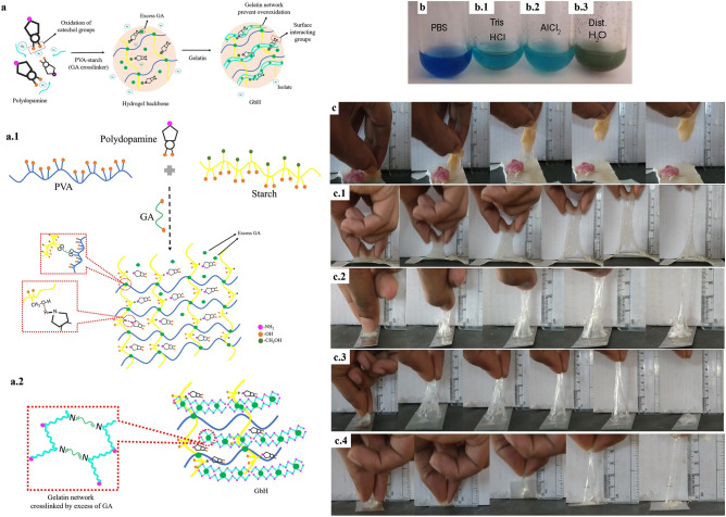
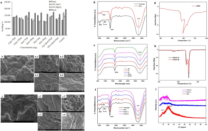
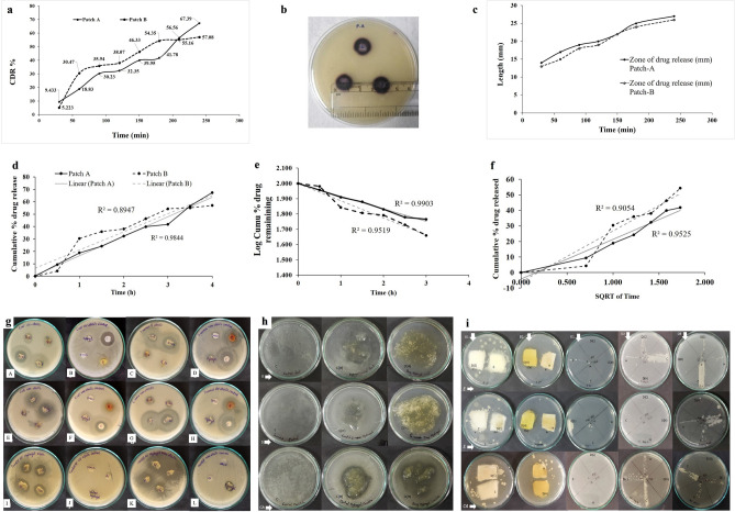
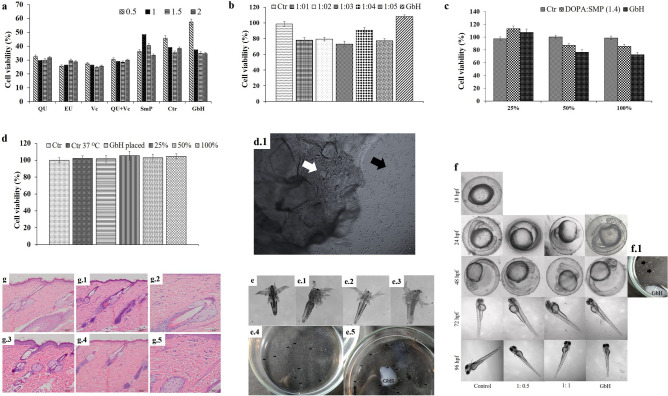
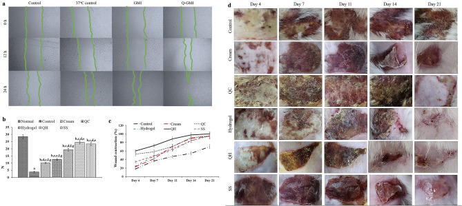
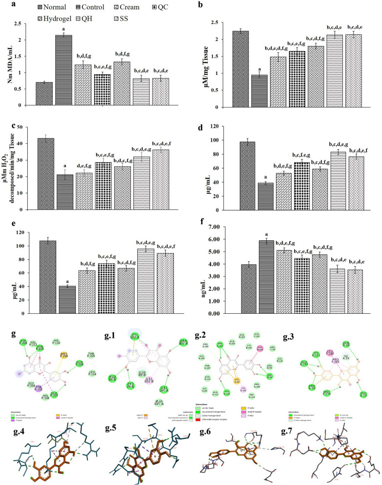
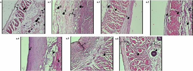
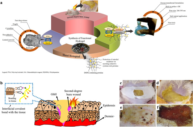
Proper burn wound management considers patient's compliance and provides an environment to accelerate wound closure. Sticky hydrogels are conducive to wound management. They can act as a preventive infection patch with controlled drug delivery and diverse surface adherence. A hypothesis-driven investigation explores a bioinspired polydopamine property in a gelatin-based hydrogel (GbH) where polyvinyl alcohol and starch function as hydrogel backbone. The GbH displayed promising physical properties with O-H group rich surface. The GbH was sticky onto dry surfaces (glass, plastic and aluminium) and wet surfaces (pork and chicken). The GbH demonstrated mathematical kinetics for a transdermal formulation, and the in vitro and in vivo toxicity of the GbH on test models confirmed the models' healthy growth and biocompatibility. The quercetin-loaded GbH showed 45-50% wound contraction on day 4 for second-degree burn wounds in rat models that were equivalent to the silver sulfadiazine treatment group. The estimates for tensile strength, biochemicals, connective tissue markers and NF-κB were restored on day 21 in the GbH treated healed wounds to imitate the normal level of the skin. The bioinspired GbH promotes efficient wound healing of second-degree burn wounds in rat models, indicating its pre-clinical applicability.


Conflict of interest statement

The authors declare no competing interests.

Figures

Figure 1
Figure 1
Schematic and diverse performance of GbH: (a) Schematic of the developed diverse surface gelatin-based hydrogel (GbH); (a.1) The developed GbH is a two-stage process where polymers of polyvinyl alcohol (PVA) and starch, and the optimized alkali polymerized polydopamine concentration is chemically crosslinked with glutaraldehyde reagent (GA), which act as the base gel; (a.2) The excess glutaraldehyde in the semi-polymerized stage crosslinks with gelatin and forms a polymer network over the base gel. The gelatin network prevents external oxygen and inhibits the oxidation of catechol groups.; The Benedict test was used to determine excess glutaraldehyde comprised: (b) PBS, (b.1) Tris–HCl, (b.2) AlCl2 and (b.3) distilled water. The developed GbH with stabilizer on diverse surfaces: (c) chicken; (c.1) pork; (c.2) stainless steel surface; (c.3) glass surface and (c.4) plastic surface.
Figure 2
Figure 2
Characterization of the GbH: The physical evaluation of GbH: (a) the swelling behaviour of GbH in water, NaCl solution, MgCl2 solution and blood. The SEM image: (b) and (c) control GbH with pore size range from 266-393 nm; (b.1) antibacterial ciprofloxacin drug-loaded GbH; (b.2) wound healing promoting quercetin drug-loaded GbH; (b.3) antifungal drug 5-flucytosine loaded GbH; (b.4) Combination of antibacterial drug ciprofloxacin and quercetin loaded GbH. (c.1) SEM image of patch-A GbH before the release of salicylic acid; (c.2) SEM image of patch-A GbH after the release of salicylic acid (c.3) SEM image of patch-B GbH before the release of salicylic acid; (c.4) SEM image of patch-B GbH after the release of salicylic acid. Surface functional characteristics illustrated: (d) FT-IR of standard hydrogel and GbH; (e) FT-IR of GbH with antibacterial drug ciprofloxacin (C) loaded, wound healing promoting drug, quercetin loaded (Q), GbH with antifungal drug 5-flucytosine (5F) loaded, GbH with antibacterial drug ciprofloxacin and quercetin combined (Q + C) loaded and GbH with antifungal drug 5-flucytosine and quercetin combined (Q + 5F) loaded; (f) FT-IR of patch-A and -B. DSC characterization of patches: (g) GbH and (h) patch-A and -B. (i) XRD of the GbH as patch-A and -B.
Figure 3
Figure 3
Mathematical modelling for transdermal drug delivery system: Time-dependent evaluation of salicylic acid release from GbH: (a) graphical illustration of spectrophotometric analysis of controlled drug release profile of patch-A and -B for salicylic acid; (b) illustration of agar well diffusion test for controlled drug release profile of patch-A and -B; (c) graphical illustration of controlled drug release profile of patch-A and -B for salicylic acid agar well diffusion test. Mathematical model representation of drug release from GbH: (d) zero-order, (e) first-order and (f) Higuchian release kinetics. Agar diffusion of GbH: (g.A) UV-sterile GbH performance against E.coli; (g.B) Control plate of UV-sterile GbH performance against E.coli; (g.C) UV-sterile GbH performance against S. aureus; (g.D) Control plate of UV-sterile GbH performance against S. aureus; (g.E) Non-sterile GbH performance against E.coli; (g.F) Control plate of non-sterile GbH performance against E.coli; (g.G) Non-sterile GbH performance against S. aureus; (g.H) Control plate of non-sterile GbH performance against S. aureus; (g.I) UV-sterile GbH performance against C. albicans; (g.J) Control plate of UV-sterile GbH performance against C. albicans; (g.K) Non-sterile GbH performance against C. albicans and (g.L) Control plate of non-sterile GbH performance against C. albicans. (h) illustrate agar overlay performance of GbH where E, S and CA denote E. coli, S. aureus and C. albicans, respectively; C denotes Control plate; C(H) denotes positive control GbH; D(H) denotes test drug-loaded GbH. (i) illustrates patch agar performance of GbH where D1, D2 and D3 are Day 1, 2 and 3, respectively; E, S and CA denote E. coli, S. aureus and C. albicans, respectively; C denotes Cotton patch; D(C) denotes drug-loaded cotton patch; H denotes GbH and D(H) denotes drug-loaded GbH.
Figure 4
Figure 4
In vitro and in vivo performance of GbH as a dressing material for second-degree burn wounds: Cytotoxicity assessment of the GbH using L929 cells: (a) Cell viability of L929 when exposed to GbH for 24 h, where Hb is hydrogel base with no stabilizer and the legend denotes the concentration (mg) of each test stabilizer; (b) Cell viability of 3T6 when exposed to the GbH leachate medium for 24 h, where Ctr is the GbH base with no stabilizer and the legend denotes the ratio of polydopamine to sodium metaperiodate and (c) Cell viability of 3T6 when exposed to the GbH leachate media for 24 h at a different percentage, where Ctr is the GbH base with no stabilizer and the legend of the ratio denotes polydopamine and sodium metaperiodate ratio in the GbH. (d) Cell viability of the HaCat when the GbH is in contact with the cells (GbH placed) and leachate medium at 100% medium replacement incubated for 24 h, where Ctr denotes control, Ctr 37 °C denotes the positive control; (d.1) Observed event of healthy HaCat cells after 24 h in contact with the GbH, where white arrow denotes the GbH and black arrow denotes healthy HaCat cells in 96-well plate. Observed development of brine shrimp when exposed to the GbH for 24 h, (e) Positive control: nauplii from the aerated tank; (e.1) Control: nauplii in artificial saltwater; (e.2) and (e.4) Test: nauplii in 1:1 medium of the GbH leeched buffer and artificial saltwater; (e.3) and (e.5) Test: The GbH placed in brine shrimp in an artificial saltwater medium. Short–term toxicity test on embryo and sac-fry stages of zebrafish model: (f) Observed development of zebrafish when exposed to the GbH (GbH direct contact and 1:1 medium, i.e., the GbH leeched buffer and E3 medium) for 18–96 hpf; (f.1) Observed healthy zebrafish eggs at 48 hpf in contact with the GbH. Photomicrographs showing the histopathology of the skin in rats. Normal appearances of the epithelium in the male (g) and female (g.1) rats of the control group. Minimal acanthosis of the male (g.3) and female (g.4) rats. Normal dermis appearance in the control rat (g.2) and minimal inflammatory cell infiltration in the treated (g.5) group.
Figure 5
Figure 5
Wound healing performance of GbH: (a) Scratch wound healing performance of HaCat cells in 100% replaced of the GbH incubated medium. (b) Changes in skin tensile strength in the Normal, Control, cream base group (Cream), quercetin loaded cream (QC), GbH base (Hydrogel), quercetin loaded GbH (QH) and silver sulfadiazine (SS) treated groups. The results are represented as the mean ± S.D. with n = 6 in each group ap < 0.05 as compared to the Normal group; bp < 0.05, as compared to Control group; cp < 0.05, as compared to Cream group, dp < 0.05, as compared to QC group, ep < 0.05, as compared to Hydrogel group, fp < 0.05, as compared to QH group, gp < 0.05, as compared to SS group. (c) The effect of treatments on burn wound contraction (%) on day 4, day 7, day 11, day 14 and day 21 was a superior and faster wound contraction compared with the control and placebo groups. (d) Changes in wound contraction of the normal, control, Cream, QC, Hydrogel, QH and SS treated groups.
Figure 6
Figure 6
In vivo wound healing changes in (a) MDA; (b) GSH; (c) CAT; (d) HXP; (e) HXA and (f) NF-κB levels of the Normal, Control, cream base group (Cream), quercetin loaded cream (QC), GbH base (Hydrogel), quercetin loaded GbH (QH) and silver sulfadiazine (SS) treated groups. The results are represented as the mean ± S.D. with n = 6 in each group ap < 0.05 as compared to the Normal group; bp < 0.05, as compared to Control group; cp < 0.05, as compared to Cream group, dp < 0.05, as compared to QC group, ep < 0.05, as compared to Hydrogel group, fp < 0.05, as compared to QH group, gp < 0.05, as compared to SS group. 2-D and 3-D image of quercetin interaction with: (g) and (g.4) 1SVC target interaction; (g.1) and (g.5) 3BRV target interaction; (g.2) and (g.6) 1NFI target interaction, and (g.3) and (g.7) 2E7A target interaction.
Figure 7
Figure 7
Burn wound histopathological assessment on day 21: Standard morphology of the epidermis, dermis and hypodermis was found during photomicrographs of a histopathological examination of the Normal group (a). The Control group (a.1) was observed to have marked characteristics of burn crust development in the dermis and epidermis, extreme leukocyte aggregation, congestion of the blood vessels, hair follicles, and the sebaceous glands. Moderate leukocytes, blood vessel destruction, and degenerating degeneration of hair follicles and sebaceous glands were observed in the topical application of the Cream and the GbH (Hydrogel) placebo group (a.2) and (a.3). The quercetin drug-treated cream and the GbH group (QC, QH) and silver sulfadiazine (SS) (a.4), (a.5) and (a.6) displayed no symptoms of leukocyte accumulation, favoured by epithelium regeneration. The restored skin architecture refelects the regeneration of the epidermis upon the topical application of quercetin loaded GbH, which is in accordance with normal skin.
Figure 8
Figure 8
Highlights of the GbH in the current research: (a) Development of bioinspired gelatin-based diverse surface sticky hydrogel for second-degree burn wound care; (b) Schematic of the surface interaction of the catechol group with the burn wound area; (c) soft wound patch interaction with wound tissue; soft wound patch; (d) QH placed on second-degree burn wound; (e) Observable wound repair on day 3 (arrow) of dressing and (f) Absorption of exudates: a hallmark of the ideal hydrogel.

References

    1. Zhao Y, et al. Supramolecular adhesive hydrogels for tissue engineering applications. Chem. Rev. 2022 doi: 10.1021/acs.chemrev.1c00815. - DOI - PubMed
    1. Li J, et al. Tough adhesives for diverse wet surfaces. Science. 2017 doi: 10.1126/science.aah6362. - DOI - PMC - PubMed
    1. Montazerian H, et al. Stretchable and bioadhesive gelatin methacryloyl-based hydrogels enabled by in situ dopamine polymerization. ACS Appl. Mater. Interfaces. 2021 doi: 10.1021/acsami.1c10048. - DOI - PubMed
    1. Han L, et al. Tough, self-healable and tissue-adhesive hydrogel with tunable multifunctionality. NPG Asia Mater. 2017 doi: 10.1038/am.2017.33. - DOI
    1. Zhang W, et al. Catechol-functionalized hydrogels: biomimetic design, adhesion mechanism, and biomedical applications. Chem. Soc. Rev. 2020 doi: 10.1039/c9cs00285e. - DOI - PMC - PubMed