Review
. 2022 Aug 12;5(1):809.
doi: 10.1038/s42003-022-03749-3.

The exquisitely preserved integument of Psittacosaurus and the scaly skin of ceratopsian dinosaurs

Affiliations
Review

The exquisitely preserved integument of Psittacosaurus and the scaly skin of ceratopsian dinosaurs

Phil R Bell et al. Commun Biol. .

Abstract

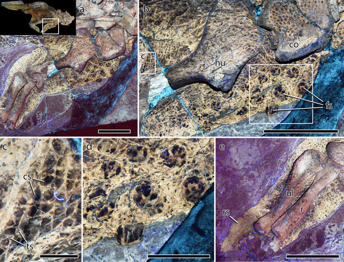
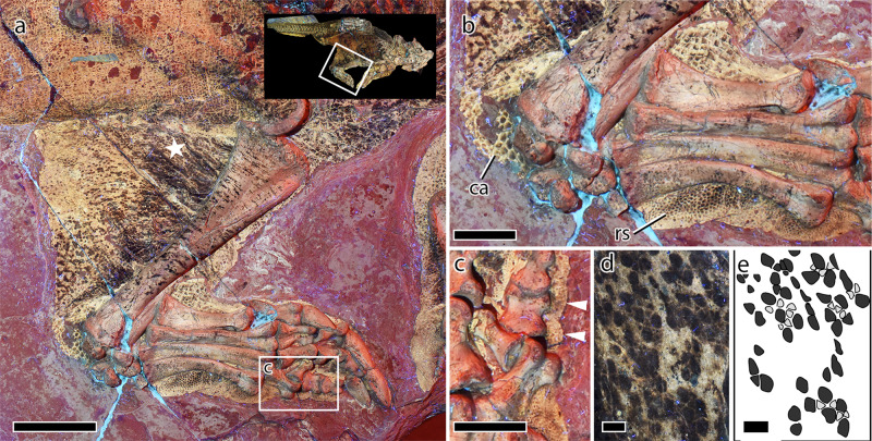
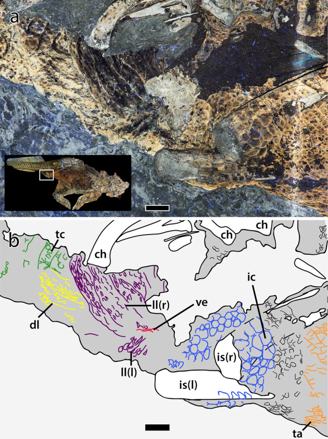
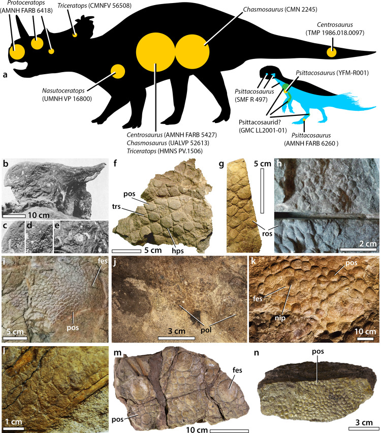
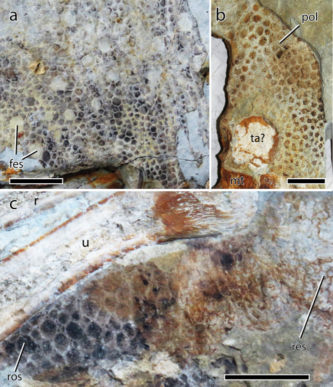
The Frankfurt specimen of the early-branching ceratopsian dinosaur Psittacosaurus is remarkable for the exquisite preservation of squamous (scaly) skin and other soft tissues that cover almost its entire body. New observations under Laser-Stimulated Fluorescence (LSF) reveal the complexity of the squamous skin of Psittacosaurus, including several unique features and details of newly detected and previously-described integumentary structures. Variations in the scaly skin are found to be strongly regionalized in Psittacosaurus. For example, feature scales consist of truncated cone-shaped scales on the shoulder, but form a longitudinal row of quadrangular scales on the tail. Re-examined through LSF, the cloaca of Psittacosaurus has a longitudinal opening, or vent; a condition that it shares only with crocodylians. This implies that the cloaca may have had crocodylian-like internal anatomy, including a single, ventrally-positioned copulatory organ. Combined with these new integumentary data, a comprehensive review of integument in ceratopsian dinosaurs reveals that scalation was generally conservative in ceratopsians and typically consisted of large subcircular-to-polygonal feature scales surrounded by a network of smaller non-overlapping polygonal basement scales. This study highlights the importance of combining exceptional specimens with modern imaging techniques, which are helping to redefine the perceived complexity of squamation in ceratopsians and other dinosaurs.


Conflict of interest statement

The authors declare no competing interests.

Figures

Fig. 1
Fig. 1. Psittacosaurus sp. (SMF R 4970) under laser-stimulated fluorescence (LSF) and distribution of different integumentary types.
a Specimen under LSF, with close ups on different integumentary types (bh). b Feature scales of the ventral portion of the tail. c Umbilical scar with transverse rows of quadrangular scales on the abdomen. d Cloaca. e Reticulate scales of the left foot. f Feature scales of the shoulder. g Jugal horn. h Basement scales of the mandible. i Schematic showing the distribution of different integumentary types. bas basement scale, bri tail bristles, clo cloaca, fes feature scale, hps hexagram pattern of basement scales, ish ischial callosity, juh jugal horn, lba large basement scales, res reticulate scales, sba small basement scales, tbs transversely banded scales, umb umbilical scar.
Fig. 2
Fig. 2. Integument of the head and neck of Psittacosaurus SMF R 4970 under LSF.
a Head and neck with inset showing the region depicted in a. b Close up of the right jugal horn. c Close up of the left jugal horn and dark region interpreted as the keratinous “horn”. Note the offset between the posterior edges of the jugal horn (black arrow) and the keratinous “horn” (black and white arrowhead). Close up on the integument of the head, from d the dorsomedial side of the left ramus of the mandible, directly ventral to the lower dentition; e the palatal region; and f the ventromedial side of the distal portion of the left ramus of the mandible. g Close up of the integument on the left side of the cervical vertebrae. h Detail of the squamous integument shown in f. i Detail of the right jugal horn (boxed region in b) with pigmented strips forming polygons (arrowheads). an angular, cv cervical vertebrae, de dentary, je jugal horn, kh keratinous “horn”, mx maxilla, qu quadrate. Scale bars equal 5 cm (a, c), 3 cm (b, d), and 5 mm (e, f).
Fig. 3
Fig. 3. Integument of the forelimb and pectoral region of Psittacosaurus SMF R 4970 under LSF.
a Left forelimb in medial aspect with inset showing the region depicted in a. b Shoulder region showing large basement scales on the coracoid, grid-like arrangement of smaller basement scales dorsal to the humerus, and large pigmented feature scales on the anterior brachium. c Detail of boxed region in b showing hexagram arrangement of basement scales. d Detail of the raised feature scales close to the shoulder joint showing striped pigmentation. e Antebrachium and fleshy palmar pad bearing reticulate scales. co coracoid, cs central scale within hexagram pattern, fes feature scales, hu humerus, r radius, res reticulate scales, ts triangular scales within hexagram pattern, ul ulna. Scale bars equal 5 cm (a, b, e), 2 cm (d), and 5 mm (c).
Fig. 4
Fig. 4. Abdominal skin in Psittacosaurus SMF R 4970 under LSF.
a Abdominal region with inset showing the region depicted in a. The maximal anteroposterior extent of the paired scales is indicated by arrowheads. b, c Close up of boxed region in a showing paired quadrangular scales (blue outline in c). Transverse banding is visible in the remaining abdominal scales (black outlines in c). d Close up of paired quadrangular scales (pqs). Scale bars equal 5 mm (ac), and 2 mm (d).
Fig. 5
Fig. 5. Hindlimb and integument of Psittacosaurus SMF R 4970 under LSF.
a Left hindlimb in medial aspect with inset showing the region depicted in a. b Close up of the left pes showing large scales forming a callosity around the ankle joint and reticulate scales covering the plantar surface of the metatarsus. c Detail of the fourth pedal digit (boxed region in a) showing undulations in the integument corresponding to arthrally arranged plantar pads (arrowheads). d, e Detail of the scales on the inner leg (star in a) showing hexagram pattern of light-colored triangular scales and darker central scales. ca callosity, res reticulates scales. Scale bars equal 5 cm (a), 2 cm (b, c), and 2 mm (d, e).
Fig. 6
Fig. 6. Cloaca of Psittacosaurus SMF R 4970 under LSF.
a Photograph and b interpretive illustration with inset showing the region depicted in a and b. Colors in b depict skeletal elements (white); integument (dark gray); lateral lips (purple) and dorsal lobe (yellow) of the cloaca; cloacal vent (red); ischial callosity (blue); transversely-banded caudal scales (green) and; transversely-banded abdominal scales (orange). ch chevron, dl dorsal lobe, ic ischial callosity, is ischium, l left (in brackets), ll lateral lip, r right (in brackets), ta transversely banded abdominal scales, tc transversely banded caudal scales, ve vent. Scale bars equal 1 cm.
Fig. 7
Fig. 7. Tail of Psittacosaurus SMF R 4970 under LSF.
a Photograph and b interpretive illustration with inset showing the region depicted in a and b. In b: integumentary outline (gray), tail bristles (blue), matrix (brown), transversely banded epidermal scales (thin black outlines), large quadrangular feature scales (green outline), dorsal pigmentation (black). Close up of the feature scales (c) and transversely banded epidermal scales (d). cv11 11th caudal vertebra, cv16 16th caudal vertebra, fes feature scales. Scale bars equal 2 cm.
Fig. 8
Fig. 8. Cloaca of Psittacosaurus and Crocodylus.
a Cloacal region of Psittacosaurus (SMF R 4970) under LSF showing the ischial callosity, transversally banded abdominal and caudal basement scales, and lateral lips of the cloaca. b Cloacal region of a juvenile freshwater crocodile (Crocodylus johnsoni; University of New England Natural History Museum, no specimen number) showing the rosette pattern of scales surrounding the vent and transverse banding of the ventral scales on the abdomen and tail. c Close up on the cloaca of Psittacosaurus (SMF R 4970) under LSF showing the two lateral lips and the longitudinally-oriented cloacal vent. d Close up on the cloaca and longitudinally-oriented vent of Crocodylus johnsoni. cl cloacal scales, dl dorsal lobe, ic ischial callosity, l left (in brackets), ll lateral lip, r right (in brackets), ta transverse abdominal scales, tc transverse caudal scales, ve vent. Anterior is toward the top in all images. Scale bars equal 2 cm (a), 1 cm (b, c), and 5 mm (d).
Fig. 9
Fig. 9. Integumentary structures in ceratopsian dinosaurs.
a Distribution of known scaly integument on the body of psittacosaurids (right silhouette; Jaime Headden; https://creativecommons.org/licenses/by/3.0/; modified) and coronosaurian ceratopsians (left silhouette; Caleb M. Brown; https://creativecommons.org/licenses/by-sa/3.0/; modified), with the position of the integument in the Psittacosaurus specimen SMF R 497, the putative psittacosaurid specimen GMC LL2001-01, and the other ceratopsian specimens in blue, green and orange, respectively. Mummified head covered with skin possibly made of minute pebbly basement scales in the protoceratopsid Protoceratops (AMNH FARB 6418; from Brown and Schlaikjer, modified) in left lateral view (b), with close up on putative basement scales of the beak (c), the lacrimal part (d), and the cheek (e). Patches of skin made of triangular, polygonal or subcircular basement scales from the proximal forearm (f, h) and shoulder region (g) of the left forelimb of the centrosaurine Nasutoceratops titusi (UMNH VP 16800; courtesy of Erik K. Lund). i Patches of skin made of feature and polygonal basement scales from the thoracic region of the centrosaurine Centrosaurus apertus (AMNH FARB 5427; courtesy of Carl Mehling). j Polygonal basement scales from the distal tail region of the centrosaurine Centrosaurus sp. (TMP 1986.018.0097; courtesy of Caleb Brown). k Polygonal feature and basement scales from the flank of the chasmosaurine Triceratops horridus (HMNS PV.1506; courtesy of Marschal A. Fazio). l Polygonal basement scales in a juvenile individual of the chasmosaurine Chasmosaurus belli (UALVP 52613; courtesy of Philip J. Currie). Two patches of skin, one with large rounded feature scales surrounded by smaller polygonal basement scales (m) and a second with small polygonal basement scales (n), from the pelvis arch and right flank of an adult specimen of the chasmosaurine Chasmosaurus belli (CMN 2245; from S. E. Pan/Canadian Museum of Nature, used under CC BY-NC 4.0; These images were cropped from the original). fes feature scale, hps hexagram pattern of basement scales, nip nipple-like structure on the feature scale, pos polygonal basement scale, ros rounded basement scale, trs triangular basement scale.
Fig. 10
Fig. 10. Integument in a possible psittacosaurid specimen (GMC LL2001-01) from the Dawangzhangzi Village of Western Liaoning, China.
a Rounded feature scales and polygonal basement scales from the shoulder and possibly neck regions. b Polygonal basement scales proximal to the metatarsals. c Polygonal basement and reticulate scales posterior to the distal half of the right ulna. fes feature scale, mt metatarsus, pol polygonal basement scale, r radius, res reticulate scale, ros rounded basement scale, ta? possible tarsus, u ulna. Scale bars equal 10 mm.
Fig. 11
Fig. 11. Basement scale length vs. body mass in non-avian dinosaurs.
Log-plot of basement scale length (taken from the largest scale) versus body mass in ceratopsians (purple dots; silhouette: Scott Hartman; https://creativecommons.org/licenses/by-nc-sa/3.0/; modified), Kulindadromeus (violet dot; silhouette: Pete Buchholz; https://creativecommons.org/licenses/by-sa/3.0/; modified), ornithopods (blue dots; silhouette: Matt Martyniuk; https://creativecommons.org/licenses/by-nc-sa/3.0/; modified), thyreophorans (yellow dots; silhouette: Scott Hartman; https://creativecommons.org/licenses/by/3.0/; modified), non-avian theropods (red squares; silhouette: Scott Hartman; https://creativecommons.org/licenses/by-nc-sa/3.0/; modified), and sauropods (green squares; silhouette: Scott Hartman; https://creativecommons.org/licenses/by-nc-sa/3.0/; modified). Ceratopsian taxa are labeled for clarity. The regression equation and r2 value are reported for ceratopsians. Figure modified from Hendrickx and Bell (ref. : Fig. 13f). Data on body masses and basement scale length is provided in the Supplementary Data.

References

    1. Mantell, G. A. On the structure of the Iguanodon, and on the fauna and flora of the Wealden Formation. Notice Proceedings, Royal Institute of Great Britain1, 141–146 (1852).
    1. Czerkas, S. A. Skin. in Encyclopedia of Dinosaurs (eds. Currie, P. J. & Padian, K.) 669–675 (Academic Press, 1997).
    1. Upchurch P, Mannion PD, Taylor MP. The anatomy and phylogenetic relationships of “Pelorosaurus“ becklesii (Neosauropoda, Macronaria) from the Early Cretaceous of England. PLoS ONE. 2015;10:e0125819. doi: 10.1371/journal.pone.0125819. - DOI - PMC - PubMed
    1. Pittman M, Enriquez NJ, Bell PR, Kaye TG, Upchurch P. Newly detected data from Haestasaurus and review of sauropod skin morphology suggests Early Jurassic origin of skin papillae. Commun. Biol. 2022;5:1–8. doi: 10.1038/s42003-022-03062-z. - DOI - PMC - PubMed
    1. Czerkas SA. The history and interpretation of sauropod skin impressions. Gaia. 1994;10:173–182.

Publication types