. 2022 Aug 12;19(1):204.
doi: 10.1186/s12974-022-02566-z.

Sensory neuron dysfunction in orthotopic mouse models of colon cancer

Affiliations

Sensory neuron dysfunction in orthotopic mouse models of colon cancer

Mihály Balogh et al. J Neuroinflammation. .

Abstract

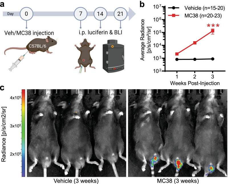
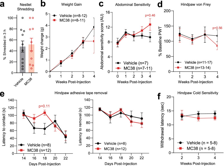
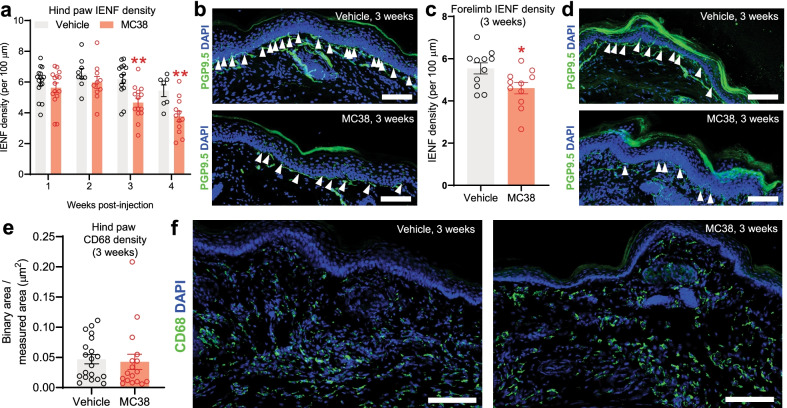
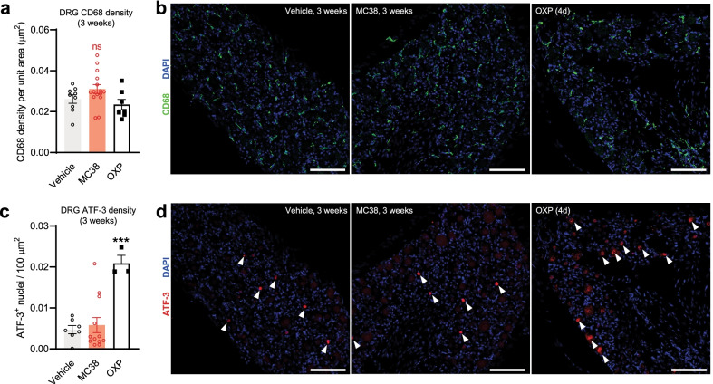
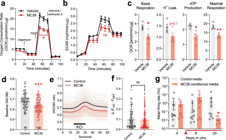
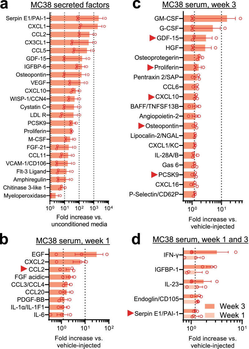
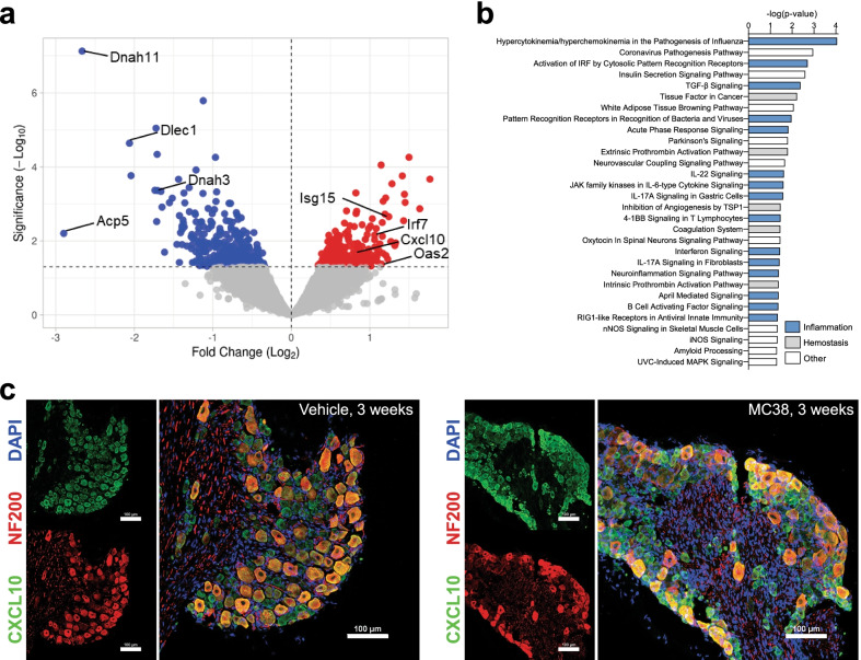
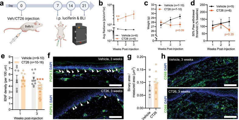
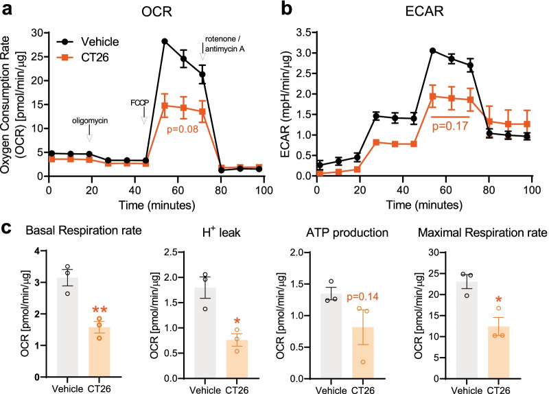
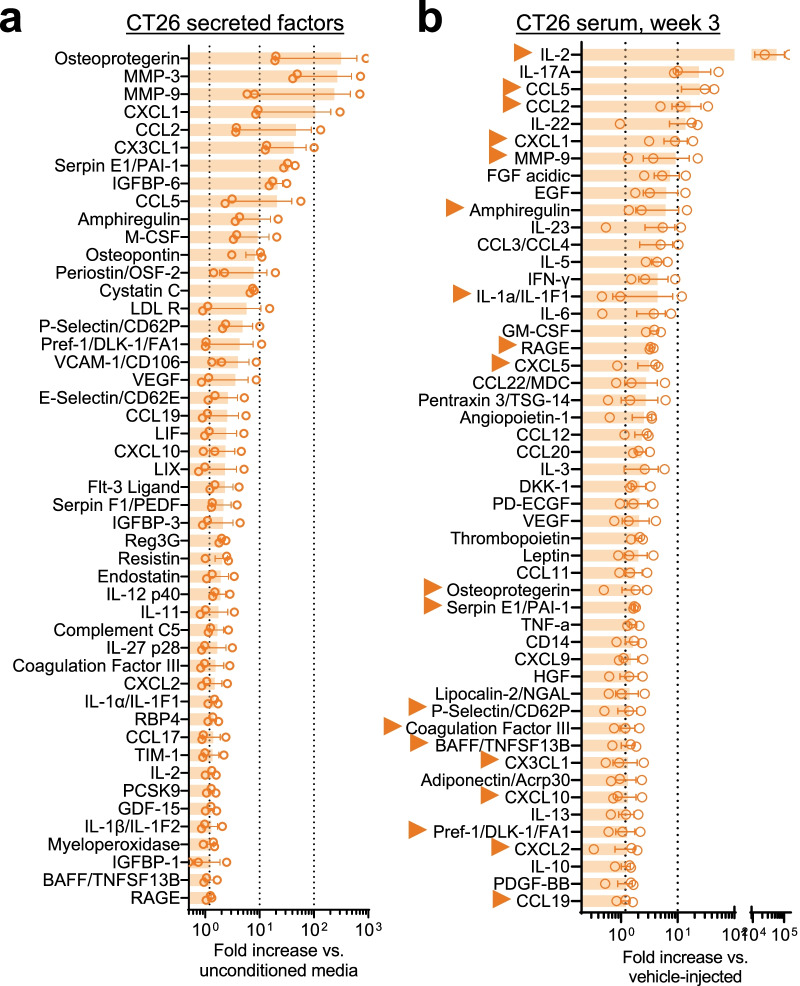
Reports of neurological sequelae related to colon cancer are largely restricted to rare instances of paraneoplastic syndromes, due to autoimmune reactions. Systemic inflammation associated with tumor development influences sensory neuron function in other disease models, though the extent to which this occurs in colorectal cancer is unknown. We induced orthotopic colorectal cancer via orthotopic injection of two colorectal cancer cell lines (MC38 and CT26) in two different mouse strains (C57BL/6 and Balb/c, respectively). Behavioral tests of pain sensitivity and activity did not detect significant alterations in sensory sensitivity or diminished well-being throughout tumor development. However, immunohistochemistry revealed widespread reductions in intraepidermal nerve fiber density in the skin of tumor-bearing mice. Though loss of nerve fiber density was not associated with increased expression of cell injury markers in dorsal root ganglia, lumbar dorsal root ganglia neurons of tumor-bearing animals showed deficits in mitochondrial function. These neurons also had reduced cytosolic calcium levels in live-cell imaging and reduced spontaneous activity in multi-electrode array analysis. Bulk RNA sequencing of DRGs from tumor-bearing mice detected activation of gene expression pathways associated with elevated cytokine and chemokine signaling, including CXCL10. This is consistent with the detection of CXCL10 (and numerous other cytokines, chemokines and growth factors) in MC38 and CT26 cell-conditioned media, and the serum of tumor-bearing mice. Our study demonstrates in a pre-clinical setting that colon cancer is associated with latent sensory neuron dysfunction and implicates cytokine/chemokine signaling in this process. These findings may have implications for determining risk factors and treatment responsiveness related to neuropathy in colorectal cancer.

Keywords: Colon cancer; DRG neuron; Neuropathic pain; Neuropathy; Paraneoplastic neuropathy.


Conflict of interest statement

The authors declare that they have no competing interests.

Figures

Fig. 1
Fig. 1
In vivo bioluminescence imaging of colon cancer development after local MC38 cell injection. a Schematic depicting of experimental protocol for IVIS bioluminescence measurements (animals were measured 7, 14, and 21 days after vehicle/MC38 cell injection). b Changes in average bioluminescence radiance values. The consistent localized increase in the measured bioluminescence signal reflects consistent tumor growth after transanal MC38 cell injection. ***: p < 0.001 vs MC38 week 1, mixed-effects model with Šídák's multiple comparisons test. c Representative image of three control (vehicle injected) and colon cancer bearing (MC38-injected) mice, 3 weeks after inoculation
Fig. 2
Fig. 2
Animal well-being- and pain-related behavior tests in the MC38 colon cancer model. a Nestlet shredding assay. Columns depict percentage (by weight) of nestlet shredded after the 3 h experimental time period. No change in the nestlet shredding indicates proper physiological behavior (p = 0.8, two-tailed unpaired t test). b Weight gain (g) of vehicle vs MC38-injected mice after inoculation (compared to baseline at week 0, before injection). Data show no significant difference in the growth of mice with or without colon cancer (mixed-effects analysis with Šídák's multiple comparisons test). c Changes in abdominal sensitivity of vehicle vs MC38-injected mice after inoculation. The graph shows the scored response provoked by the application of the 0.02 g Von Frey filament to the abdomen (mixed-effects analysis with Šídák's multiple comparisons test). d Mechanical sensitivity of hind paws, as measured by the application of von Frey filaments, by the up-and-down method. Datapoints represent the average of percentage changes, as compared to the measured individual baseline values (measured at week 0, before inoculation). Animals did not develop significant mechanical allodynia (mixed-effects analysis with Šídák's multiple comparisons test). e Changes in the removal of adhesive tape from the plantar surface of the hindpaw, depicted as latency to contact (s), and latency to removal (s), respectively. Animals with colon cancer did not develop elevated or diminished tactile sensitivity (mixed-effects analysis with Šídák's multiple comparisons test). f Cold sensitivity of hind paws. Withdrawal threshold: time until sudden removal of the hind paw (s). The induction of colon cancer did not result in elevated cold sensitivity in the experimental period (mixed-effects analysis with Šídák's multiple comparisons test)
Fig. 3
Fig. 3
IHC assessment of hind paw and forelimb samples after MC38 injection. a Changes in IENF density with time in the hind paws of control vs MC38-injected mice. Colon cancer initiates a significant decrease in IENF density after the 3rd week, indicating significant neuronal damage. (**: p < 0.01; n = 9–13/group, two-way ANOVA with Šídák's multiple comparisons test). b Representative images of hind paw samples, collected 3 weeks after inoculation. White arrows mark the counted intraepidermal nerve fibers, stained with PGP9.5 antibody (green). Nuclei are stained with DAPI (blue). Scale bar: 100 μm c Changes in IENF density, 3 weeks after inoculation, in the forelimb samples of control vs MC38-injected mice. Similarly to the hind paw samples, our data reveal a significant decrease in IENF density, 3 weeks after local MC38 cell injection (two-tailed unpaired t test). d Representative images of forelimb samples, collected 3 weeks after tumor inoculation. White arrows mark the counted intraepidermal nerve fibers marked by PGP9.5 antibody. Nuclei are stained with DAPI. e Signal intensity of the CD68 macrophage marker in the skin of control vs MC38-injected mice, depicted as binary area/measured area. Based on the CD68 marker, there is no significant change in the number of macrophages in the epidermis, around the intraepidermal nerve fibers (p = 0.75, two-tailed unpaired t test). f Representative images of hind paw samples show no change in the CD68 signal intensity of the epidermis. Nuclei are stained with DAPI. Scale bar: 100 μm
Fig. 4
Fig. 4
IHC assessment of the neuronal damage marker ATF-3 in DRGs. Lumbar DRG samples were collected 3 weeks after tumor inoculation. Naïve mice were treated with oxaliplatin (OXP) as a positive control for ATF-3 activation. a, b No change in the signal intensity of the macrophage marker CD68 (vehicle vs MC38-injected or oxaliplatin-treated mice). c, d Representative images show no change in ATF-3 signal (samples from a vehicle vs an MC38-injected mouse, collected 3 weeks after injection). However, oxaliplatin treatment alone (positive control; cumulative dose: 12 mg/kg i.p, in 3 days) causes neuronal damage, hence significantly elevating ATF-3 signal density. White arrows indicate cells positive for ATF-3. Scale bar: 100 μm. *** = p < 0.001, one-way ANOVA, Tukey’s multiple comparisons test
Fig. 5
Fig. 5
Mitochondrial function, Ca2+ homeostasis and spontaneous activity of lumbar DRG neurons. ac Seahorse mitochondrial function assay on DRGs collected 3 weeks after inoculation (n = 3 biological replicates/group). All values are normalized to mitochondrial-dependent respiration and the protein load. a, b Basal oxygen consumption rate (OCR) and extracellular acidification rate (ECAR) are measured prior to treatment with oligomycin (complex V inhibitor) to calculate proton leak and ATP production (c). Maximal respiration is measured post-addition of the uncoupler FCCP. Values post-rotenone/antimycin A (complex I/III inhibitors) reflect non-mitochondrial oxygen consumption. This ‘background’ oxygen consumption is subtracted from the values in c. *: p < 0.05; **: p < 0.01, two-tailed unpaired t test. d, e Live-cell Ca2+ imaging on DRGs collected 3 weeks after MC38 inoculation. Number of measured cells: n = 78 and n = 138 for the vehicle and MC38 groups, respectively, from 2 biological replicates/group. Lumbar and non-lumbar DRG neurons were cultured and tested separately for extra control (additional Fig. 1). d DRGs from MC38-injected mice show significantly lower resting/baseline [Ca2+]i compared to vehicle-injected mice. The columns depict the average values of the 60-s baseline measurements for each group (***: p < 0.001, two-tailed unpaired t test). e Traces (average traces in bold) showing changes in intracellular [Ca2 +]i after superfusion of 25 mM potassium chloride (KCl) for 15 s. f No significant change in F340:F380 amplitude induced by KCl in e (two-tailed unpaired t test). g Multielectrode array analysis of DRG neurons from naïve mice with or without the addition of MC38-conditioned media (n = 8–10 wells/group, from at least 3 different cell cultures). Neuronal activity was measured every 4 h, for 24 h. Conditioned media or control solution (unconditioned MC38 media) was added after the 4 h measurement (marked with a red arrow and line). The addition of MC38-conditioned media was associated with a significant decrease in the spontaneous activity of DRG neurons at 20 h in vitro (*: p < 0.05 vs control, Mann–Whitney U test)
Fig. 6
Fig. 6
Factors secreted by MC38 cells and inflammatory changes in MC38 tumor-bearing mice. The change in different inflammatory mediators was measured by a complex proteome profiler array and presented as fold-change in signal intensity compared to the average of given control values (n = 3/group, in duplicates). a Inflammatory mediators found to be secreted by MC38 cells (factors with elevated signal intensity in conditioned media vs un-conditioned media, as presented by fold-increase). bd Elevated inflammatory mediators in the plasma samples of MC38-injected mice compared to control (vehicle injected) mice. b Increased factors in blood samples collected 1 week after MC38 inoculation. c Increased factors in blood samples collected 3 weeks after MC38 inoculation. d Factors increased both 1 and 3 weeks after tumor cell injection. Red arrows mark mediators found to be secreted by MC38 cells, as well as increased in the collected plasma samples
Fig. 7
Fig. 7
Bulk RNA sequencing of DRGs from MC38-injected mice. Bulk RNA sequencing analysis on lumbar DRGs of mice collected 3 weeks after tumor inoculation (n = 3/group). a Volcano plot showing the different genes with significantly decreased (blue) or increased (red) expression (p≤ 0.05). b Gene expression pathway enrichment analysis (MC38-injected vs vehicle-injected mice). Blue: immune/inflammation-related pathways; grey: hemostasis/clotting/thrombotic pathways (consistent with elevation of serpin E1 and chitinase-3 like 1, among others, in the proteome profiler arrays). c Immunohistochemistry of lumbar DRG from vehicle-injected and MC38 tumor-bearing mice at 3 weeks indicates CXCL10 immunoreactivity (green) overlaps with the neuronal marker NF200 (red), as well as a subpopulation of small–medium diameter neurons. DAPI: blue, scale bar = 100 μm
Fig. 8
Fig. 8
Tumor development, behavioral-, and IENF density changes in the CT26 colon cancer model. The data from animals with local injection of CT26 cells reveal very similar changes and patterns to the MC38 model. a Schematic representation of the experimental protocol for the IVIS in vivo bioluminescence measurements (animals were measured 7, 14, and 21 days after vehicle/CT26 cell injection). The consistent robust increase in the bioluminescence signal is depicted in b and indicates persistent colon cancer development (**: p < 0.01 vs vehicle-treated, two-way ANOVA with Šídák's multiple comparisons test). c No significant change in the development (animal weight, g) of mice with or without CT26 initiated colon cancer (mixed-effects analysis with Šídák's multiple comparisons test). d Von Frey testing of hind paws revealed no significant alteration in mechanical sensitivity. Datapoints represent the average of percentage changes, compared to the measured individual baseline values (measured at week 0, before inoculation. Two-way ANOVA with Šídák's multiple comparisons test). e–h Immunohistochemical analysis of hind paw samples: changes in IENF (PGP9.5) and macrophage (CD68) signals. e Changes in IENF density with time in hind paws of control vs CT26-injected mice. The IENF density in the hind paws of CT26-injected mice was significantly decreased compared to control (vehicle inj.), 3 weeks after tumor inoculation (***: p < 0.001, two-way ANOVA with Šídák's multiple comparisons test). f Representative images show the decrease in IENF density compared to control, 3 weeks after tumor inoculation. White arrows indicate intraepidermal nerve fibers. Scale bar: 100 μm. g No significant changes in CD68 signal intensity at the 3rd week in the hind paws of control vs CT26-injected mice (p = 0.73, two-tailed unpaired t test). h Representative images of hind paw samples show no change in the CD68 signal intensity of the epidermis (3 weeks after cell injection). Scale bar: 100 μm
Fig. 9
Fig. 9
Seahorse assay of DRG cells in the CT26 model. The mitochondrial function assay—performed on living lumbar DRGs of control and colon cancer bearing mice—reveals significant mitochondrial dysfunction 3 weeks after CT26 cell injection (similarly to the MC38 model). All values are normalized to mitochondrial-dependent respiration and the protein load. a, b Trend to reduced OCR and ECAR after addition of the uncoupler FCCP. c Deficits in mitochondrial function in CT26 versus vehicle-injected mice become statistically significant when corrected for non-mitochondrial oxygen consumption. *: p < 0.05; **: p < 0.01, two-tailed unpaired t test
Fig. 10
Fig. 10
Factors secreted by CT26 cells and inflammatory changes in CT26 tumor-bearing mice. The change in different inflammatory mediators was measured by proteome profiler array and presented as fold-change in signal intensity compared to mean control values (n = 3/group, in duplicates). a Inflammatory mediators found to be secreted by CT26 cells (factors with elevated signal intensity in conditioned media vs un-conditioned media, as presented by fold-increase). b Increased factors in blood samples collected 3 weeks after CT26 inoculation. Arrows mark mediators detected in CT26-injected mouse serum that were also detected in CT26-conditioned media (a)

References

    1. Kuipers EJ, Grady WM, Lieberman D, Seufferlein T, Sung JJ, Boelens PG, et al. Colorectal cancer. Nat Rev Dis Prim. 2015;1:15065. doi: 10.1038/nrdp.2015.65. - DOI - PMC - PubMed
    1. Naghavi M, Wang H, Lozano R, Davis A, Liang X, Zhou M, et al. Global, regional, and national age-sex specific all-cause and cause-specific mortality for 240 causes of death, 1990–2013: a systematic analysis for the Global Burden of Disease Study 2013. Lancet. 2015;385:117–171. doi: 10.1016/S0140-6736(14)61682-2. - DOI - PMC - PubMed
    1. Siegel RL, Miller KD, Fuchs HE, Jemal A. Cancer statistics, 2021. CA Cancer J Clin. 2021;71:7–33. doi: 10.3322/caac.21654. - DOI - PubMed
    1. Bouchenaki H, Danigo A, Sturtz F, Hajj R, Magy L, Demiot C. An overview of ongoing clinical trials assessing pharmacological therapeutic strategies to manage chemotherapy-induced peripheral neuropathy, based on preclinical studies in rodent models. Fundam Clin Pharmacol. 2021 doi: 10.1111/fcp.12617. - DOI - PubMed
    1. Chung JW, Cho YH, Ahn MJ, Lee MJ, Kim GM, Chung CS, et al. Association of cancer cell type and extracellular vesicles with coagulopathy in patients with lung cancer and stroke. Stroke. 2018;49:1282–1285. doi: 10.1161/STROKEAHA.118.020995. - DOI - PubMed