. 2022 Aug 14;56(3):326-335.
doi: 10.2478/raon-2022-0028.

Nanosecond electric pulses are equally effective in electrochemotherapy with cisplatin as microsecond pulses

Affiliations

Nanosecond electric pulses are equally effective in electrochemotherapy with cisplatin as microsecond pulses

Angelika Vizintin et al. Radiol Oncol. .

Abstract

Background: Nanosecond electric pulses showed promising results in electrochemotherapy, but the underlying mechanisms of action are still unexplored. The aim of this work was to correlate cellular cisplatin amount with cell survival of cells electroporated with nanosecond or standardly used 8 × 100 μs pulses and to investigate the effects of electric pulses on cisplatin structure.

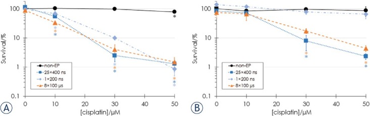
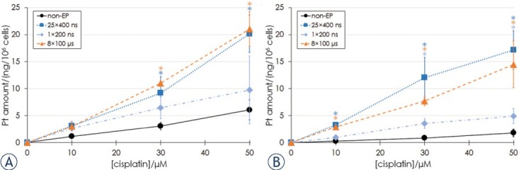
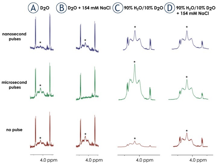
Materials and methods: Chinese hamster ovary CHO and mouse melanoma B16F1 cells were exposed to 1 × 200 ns pulse at 12.6 kV/cm or 25 × 400 ns pulses at 3.9 kV/cm, 10 Hz repetition rate or 8 × 100 μs pulses at 1.1 (CHO) or 0.9 (B16F1) kV/cm, 1 Hz repetition rate at three cisplatin concentrations. Cell survival was determined by the clonogenic assay, cellular platinum was measured by inductively coupled plasma mass spectrometry. Effects on the structure of cisplatin were investigated by nuclear magnetic resonance spectroscopy and high-resolution mass spectrometry.

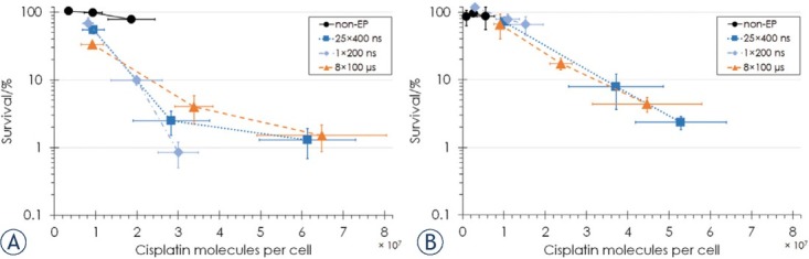
Results: Nanosecond pulses equivalent to 8 × 100 μs pulses were established in vitro based on membrane permeabilization and cell survival. Equivalent nanosecond pulses were equally efficient in decreasing the cell survival and accumulating cisplatin intracellularly as 8 × 100 μs pulses after electrochemotherapy. The number of intracellular cisplatin molecules strongly correlates with cell survival for B16F1 cells, but less for CHO cells, implying the possible involvement of other mechanisms in electrochemotherapy. The high-voltage electric pulses did not alter the structure of cisplatin.

Conclusions: Equivalent nanosecond pulses are equally effective in electrochemotherapy as standardly used 8 × 100 μs pulses.

Keywords: cisplatin; electrochemotherapy; electroporation; nanosecond pulses.


Figures

Figure 1
Figure 1
Cell survival of (A) CHO and (B) B16F1 cells at different cisplatin concentrations determined by the clonogenic assay for non-electroporated (non-EP) cells (black circles) and cells electroporated with 25 x 400 ns pulses at 3.9 kV/cm, 10 Hz repetition rate (dark blue squares), 1 × 200 ns pulse at 12.6 kV/cm (light blue diamonds) or 8 × 100 μs pulses at 1.1 (CHO) or 0.9 (B16F1) kV/cm, 1 Hz pulse repetition rate (orange triangles). Bars represent standard deviation, asterisks (*) show statistically significant differences (p < 0.05) to the survival of non-electroporated cells without cisplatin. Survival data were combined from the previous8 (for non-electroporated cells and cells electroporated with 25 × 400 ns and 8 × 100 μs pulses) and the present study (for B16F1 cells, additional non-electroporated CHO cells and CHO cells electroporated with 1 × 200 ns pulse).
Figure 2
Figure 2
Pt amount in cell pellets of (A) CHO and (B) B16F1 cells after 25 min incubation at different extracellular cisplatin concentrations in non-electroporated (non-EP) cells (black circles) and cells electroporated with 25 x 400 ns pulses at 3.9 kV/ cm, 10 Hz repetition rate (dark blue squares), 1 × 200 ns pulse at 12.6 kV/cm (light blue diamonds) or 8 × 100 μs pulses at 1.1 (CHO) or 0.9 (B16F1) kV/cm, 1 Hz pulse repetition rate (orange triangles). Bars represent standard deviation, asterisks (*) show statistically significant differences (p < 0.05) to the measured number of cisplatin molecules in non-electroporated cells at the same extracellular cisplatin concentration.
Figure 3
Figure 3
Cell survival as a function of the number of cisplatin molecules per cell for (A) CHO cells and (B) B16F1 cells in non-electroporated (non-EP) cells (black circles) and cells electroporated with 25 x 400 ns pulses at 3.9 kV/cm, 10 Hz repetition rate (dark blue squares), 1 × 200 ns pulse at 12.6 kV/cm (light blue diamonds) or 8 × 100 μs pulses at 1.1 (CHO) or 0.9 (B16F1) kV/cm, 1 Hz pulse repetition rate (orange triangles). Bars represent standard deviation. Survival data were combined from the previous (for non-electroporated CHO cells and CHO cells electroporated with 25 × 400 ns and 8 × 100 μs pulses) and the present study (for B16F1 cells, additional non-electroporated CHO cells and CHO cells electroporated with 1 × 200 ns pulse).
Figure 4
Figure 4
1H NMR spectra of cisplatin, showing the signals for hydrogens of NH3 ligands labeled with asterisks (*). Spectra were recorded in a) D2O, b) D2O containing 154 mM NaCl, c) 90% H2O/10% D2O and d) 90% H2O/10% D2O containing 154 mM NaCl treated with 25 × 400 ns pulses (blue), 8 × 100 μs pulses (green) or no pulses (red).
Figure 5
Figure 5
The mechanism of cisplatin uptake into cells is not completely elucidated. In non-electroporated cells, cisplatin enters partially through passive diffusion and facilitated diffusion through ion channels including LRRC8 volume-regulated anion channels (VRAC) and membrane transporters like copper transporter 1 (CTR1) and organic cation transporters (OCTs). In electroporated cells, more cisplatin can enter through the permeabilized cell membrane (pore is a symbolic presentation of increased membrane permeability even though the mechanisms behind electroporation are more complex – refer to34).

Similar articles

Cited by

References

    1. Miklavčič D, Mali B, Kos B, Heller R, Serša G. Electrochemotherapy: from the drawing board into medical practice. BioMed Eng Online. 2014;13:1–20. doi: 10.1186/1475-925X-13-29. - DOI - PMC - PubMed
    1. Campana LG, Miklavčič D, Bertino G, Marconato R, Valpione S, Imarisio I. Electrochemotherapy of superficial tumors – Current status: basic principles, operating procedures, shared indications, and emerging applications. Semin Oncol. 2019;46:173–91. doi: 10.1053/j.seminoncol.2019.04.002. M, et al. - DOI - PubMed
    1. Gehl J, Sersa G, Matthiessen LW, Muir T, Soden D, Occhini A, Quaglino P. Updated standard operating procedures for electrochemotherapy of cutaneous tumours and skin metastases. Acta Oncol. 2018;57:874–82. doi: 10.1080/0284186X.2018.1454602. et al. - DOI - PubMed
    1. Kendler M, Micheluzzi M, Wetzig T, Simon JC. Electrochemotherapy under tumescent local anesthesia for the treatment of cutaneous metastases. Dermatologic Surg. 2013;39:1023–32. doi: 10.1111/dsu.12190. - DOI - PubMed
    1. Schoenbach KH, Beebe SJ, Buescher ES. Intracellular effect of ultrashort electrical pulses. Bioelectromagnetics. 2001;22:440–8. doi: 10.1002/bem.71. - DOI - PubMed

Publication types

LinkOut - more resources