. 2022 Aug 16;40(7):111220.
doi: 10.1016/j.celrep.2022.111220. Epub 2022 Aug 3.

SARS-CoV-2 spike N-terminal domain modulates TMPRSS2-dependent viral entry and fusogenicity

Collaborators, Affiliations

SARS-CoV-2 spike N-terminal domain modulates TMPRSS2-dependent viral entry and fusogenicity

Bo Meng et al. Cell Rep. .

Abstract

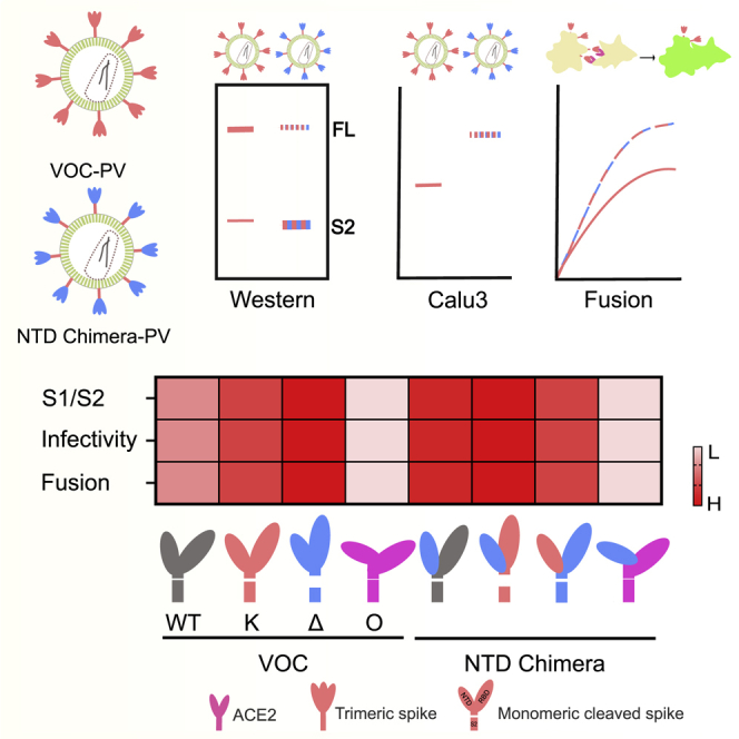
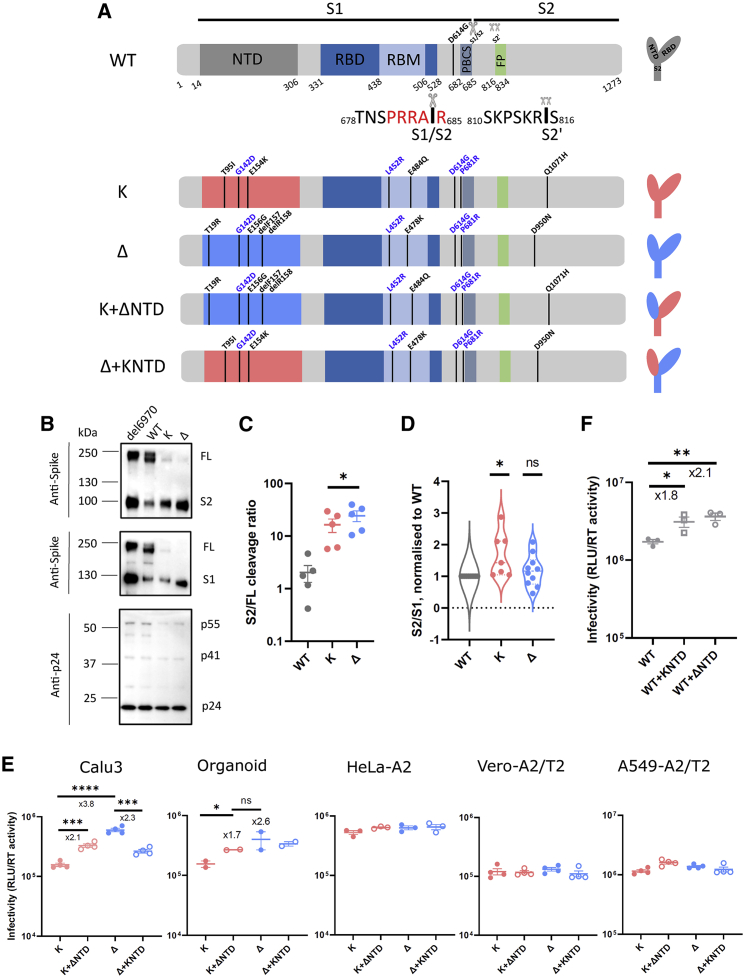
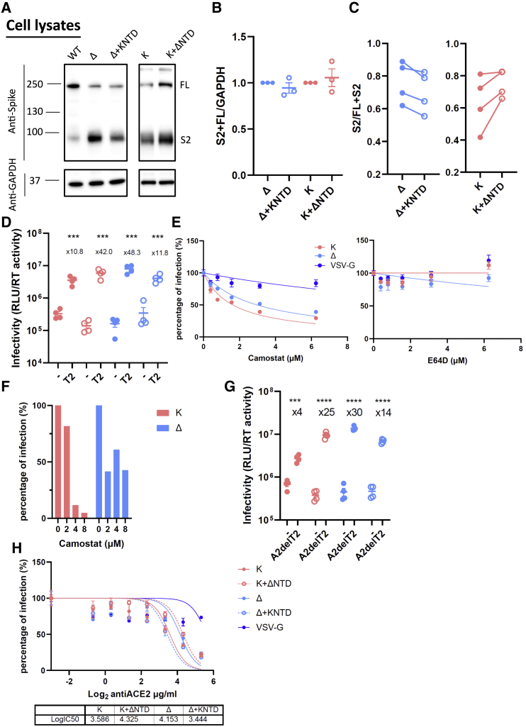
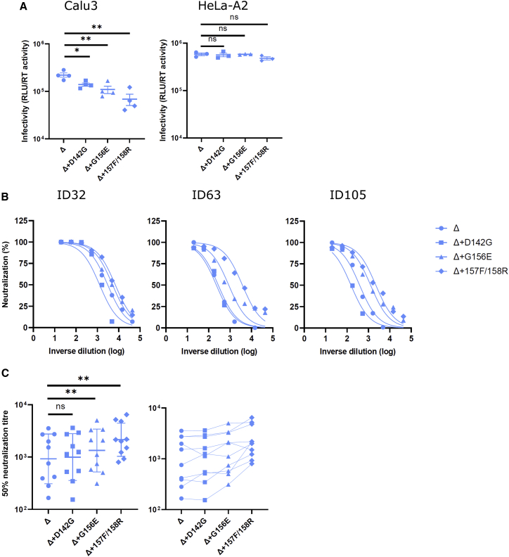
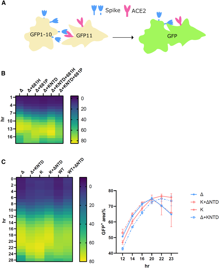
The severe acute respiratory syndrome coronavirus 2 (SARS-CoV-2) spike N-terminal domain (NTD) remains poorly characterized despite enrichment of mutations in this region across variants of concern (VOCs). Here, we examine the contribution of the NTD to infection and cell-cell fusion by constructing chimeric spikes bearing B.1.617 lineage (Delta and Kappa variants) NTDs and generating spike pseudotyped lentivirus. We find that the Delta NTD on a Kappa or wild-type (WT) background increases S1/S2 cleavage efficiency and virus entry, specifically in lung cells and airway organoids, through use of TMPRSS2. Delta exhibits increased cell-cell fusogenicity that could be conferred to WT and Kappa spikes by Delta NTD transfer. However, chimeras of Omicron BA.1 and BA.2 spikes with a Delta NTD do not show more efficient TMPRSS2 use or fusogenicity. We conclude that the NTD allosterically modulates S1/S2 cleavage and spike-mediated functions in a spike context-dependent manner, and allosteric interactions may be lost when combining regions from more distantly related VOCs.

Keywords: CP: Microbiology; Delta; NTD; Omicron; Organoid; SARS-CoV-2; TMPRSS2; entry; fusogenicity; spike.


Conflict of interest statement

Declaration of interests R.K.G. has received honoraria for consulting and educational activities from Gilead, GSK, Janssen, and Moderna. All the other authors declare no competing interests.

Figures

None
Graphical abstract
Figure 1
Figure 1
SARS-CoV-2 Delta exhibits increased infectivity over Kappa in Calu3 cells and is dependent on the NTD (A) Schematic diagrams of WT (with D614G), Kappa, and Delta with their chimeras bearing swapped NTDs. The consensus mutations between Kappa and Delta are annotated in blue. The monomeric spikes shown on the right-hand side are for illustration purposes. PBCS, polybasic cleavage site; RBM, receptor-binding motif; FP, fusion peptide. (B) Western blots of purified PVs bearing either H69V70 deletion or WT, Kappa, or Delta spikes. The sizes of protein markers are labeled to the left of the blot, and the corresponding bands are labeled to the right. (C and D) The intensity of the spike-associated bands on the western blots was densitometrically quantified (ImageJ) before the ratio was calculated for cleavage (C; S2/FL, paired t test) or spike stability (D; S2/S1; one sample t test). In both (C) and (D), each dot represents one PV preparation. (E) PV bearing Delta, Kappa, or chimeric spike was used to transduce Calu3 and organoids expressing endogenous levels of ACE2 and TMPRSS2 and ACE2/TMPRSS2-overexpressing cell lines including HeLa-ACE2, Vero-ACE2/TMPRSS2, and A549-ACE2/TMPRSS2. Unpaired t test. (F) PV bearing WT, WT with Kappa NTD, and WT with Delta NTD were used to transduce Calu3 cells. In (E) and (F), mean ± SEM are shown for technical replicates (n = 2–4; two-sided unpaired Student t test). Data are representative of two to four experiments. ns, not significant, p < 0.05, ∗∗p < 0.01, ∗∗∗p < 0.001, ∗∗∗∗p < 0.0001.
Figure 2
Figure 2
The NTD modulates the usage of TMPRSS2 and ACE2 for SARS-CoV-2 virus entry (A) A representative western blot of transfected 293T cell lysates showing spike cleavage in Delta and Kappa and their chimeras. GAPDH was probed as a loading control. (B) The band intensity from (A) was densitometrically calculated using ImageJ. Total S2-associated spike proteins (S2 and FL) were then normalized against GAPDH across Delta, Kappa, and NTD-bearing chimeras. (C) The ratio of S2/FL+S2 from the band intensity of S2 and FL shown in (A) was plotted. n ≥ 3. (D) Delta, Kappa, or chimeras was transduced into either parental 293T cells or 293T cells overexpressing TMPRSS2. The fold increase of the virus entry in T2-overexpressing cells over parental cells is shown above the scatterplots. Mean ± SEM are shown for technical replicates (n = 4; two-sided unpaired Student’s t test). (E) The entry efficiency of Delta and Kappa in A549-ACE2/TMPRSS2 cells in the presence of TMPRSS2-specific inhibitor camostat or cathepsin-specific inhibitor E64D. The lentivirus pseudotyped with VSV-G was used as a control. The relative light unit (RLU) was normalized with non-drug-added control, giving a percentage of infection. The data showing the SEM from 4 experiments were plotted; the error bars that lie within the datum points are not shown. (F) The entry efficiency of Delta and Kappa in the presence of camostat in airway organoids. (G) Delta, Kappa, or chimeras was transduced into either parental 293T cells or 293T cells overexpressing ACE2 with abrogated TMPRSS2 expression (A2delT2). The fold increase of the virus entry in A2delT2 cells over parental cells is shown above the scatterplots. Mean ± SEM are shown for technical replicates (n = 4, two-sided unpaired Student’s t test). (H) 293T-A2delT2 cells were pretreated with anti-ACE2 antibody before the addition of PV bearing either Delta, Kappa, or chimeras together with virus pseudotyped with VSV-G. The data show the SEM from 2 technical replicates. In (D) and (F)–(H), data are representative of two experiments. ∗∗∗p < 0.001, ∗∗∗∗p < 0.0001.
Figure 3
Figure 3
Reverting mutations in the Delta NTD toward WT reduces infectivity in lung cells and increases neutralization sensitivity to vaccine-elicited antibody (A) PV bearing Delta and its reversions were used to transduce Calu3 and HeLa-ACE2 cells. Mean ± SEM are shown for technical replicates (n = 4; two-sided unpaired Student’s t test). (B) Examples of neutralization curves from ID32, -63, and -105 vaccinees with PVs bearing the reversion at 142, 156, or 157/8. Data points represent the mean of two technical replicates. (C) The 50% serum neutralization was plotted across ten sera showing the geometric mean with geometric SD. Paired Wilcoxon was used for analysis. Data are representative of two experiments. ns, not significant, p < 0.05, ∗∗p < 0.01.
Figure 4
Figure 4
The SARS-CoV-2 Delta NTD increases fusion kinetics of Kappa and WT spikes (A) A schematic diagram showing the split GFP system for spike-ACE2-mediated cell fusion. (B) 681R or 681H is required for the enhanced fusogenicity in Delta and its chimera bearing the Kappa NTD. (C) The fused Delta NTD in Kappa and WT increased the fusion kinetics of their counterparts, respectively. The line graphs on the right show the percentage of the positive GFP area at 12, 14, 16, 20, 22, and 23 h post transfection. The data showing the SEM at each time point were averaged from two experiments. The heatmap at each time point shows the mean of the GFP-positive area over the field of view from two experiments.
Figure 5
Figure 5
The SARS-CoV-2 Delta NTD or BA.2 NTD does not alter spike fusion or sensitivity to TMPRSS2 of BA.1 (A) PV bearing Delta, BA.1, BA.2, or chimeric forms of BA.1 and BA.2 spike were used to transduce Calu3, H1299, and 293T expressing endogenous levels of ACE2 and TMPRSS2 and TMPRSS2-overexpressing 293T cells. (B) Fusion kinetics of the chimeric Delta NTD in BA.1 and BA.2 along with their parental spikes. The heatmap at each time point shows the mean of the GFP-positive area over the field of view from two experiments. The western blot showing cleavage of spike is directly underneath the heatmap. (C) PV bearing BA.1, BA.2, or chimeras with Delta were transduced into either parental 293T cells or 293T cells overexpressing TMPRSS2. The fold increase of the virus entry in TMPRSS2-overexpressing cells over parental cells is shown above the scatterplots. (D) The fusion assay of BA.1, BA.2, and their chimeric BA.1 bearing the BA.2 NTD and BA.2 bearing the BA.1 NTD. The line graphs show the percentage of the positive GFP area at 1 h interval post transfection. The data showing the SEM at each time point were averaged from two experiments. (E) BA.1, BA.2, or chimeras bearing the BA.2 NTD or the BA.1 NTD together with Delta were transduced into either parental 293T cells or 293T cells overexpressing TMPRSS2. The fold increase of the virus entry in T2-overexpressing cells over parental cells is shown above the scatterplots. In (A), (C), and (E), the plots are representative of two experiments. Mean ± SEM are shown for technical replicates (n = 4; two-sided unpaired Student’s t test). p < 0.05, ∗∗p < 0.01, ∗∗∗p < 0.001, ∗∗∗∗p < 0.0001.

References

    1. Barton M.I., MacGowan S.A., Kutuzov M.A., Dushek O., Barton G.J., van der Merwe P.A. Effects of common mutations in the SARS-CoV-2 Spike RBD and its ligand, the human ACE2 receptor on binding affinity and kinetics. Elife. 2021;10:e70658. doi: 10.7554/eLife.70658. - DOI - PMC - PubMed
    1. Belouzard S., Chu V.C., Whittaker G.R. Activation of the SARS coronavirus spike protein via sequential proteolytic cleavage at two distinct sites. Proc. Natl. Acad. Sci. USA. 2009;106:5871–5876. doi: 10.1073/pnas.0809524106. - DOI - PMC - PubMed
    1. Bosch B.J., Bartelink W., Rottier P.J.M. Cathepsin L functionally cleaves the severe acute respiratory syndrome coronavirus class I fusion protein upstream of rather than adjacent to the fusion peptide. J. Virol. 2008;82:8887–8890. doi: 10.1128/JVI.00415-08. - DOI - PMC - PubMed
    1. Braga L., Ali H., Secco I., Chiavacci E., Neves G., Goldhill D., Penn R., Jimenez-Guardeño J.M., Ortega-Prieto A.M., Bussani R., et al. Drugs that inhibit TMEM16 proteins block SARS-CoV-2 spike-induced syncytia. Nature. 2021;594:88–93. doi: 10.1038/s41586-021-03491-6. - DOI - PMC - PubMed
    1. Buchrieser J., Dufloo J., Hubert M., Monel B., Planas D., Rajah M.M., Planchais C., Porrot F., Guivel-Benhassine F., van der Werf S., et al. Syncytia formation by SARS-CoV-2-infected cells. EMBO. J. 2021;40:e107405. doi: 10.15252/embj.2020107405. - DOI - PMC - PubMed

Publication types

Substances

Supplementary concepts