. 2022 Oct:350:256-270.
doi: 10.1016/j.jconrel.2022.08.009. Epub 2022 Aug 24.

A lipid nanoparticle platform for mRNA delivery through repurposing of cationic amphiphilic drugs

Affiliations

A lipid nanoparticle platform for mRNA delivery through repurposing of cationic amphiphilic drugs

Bram Bogaert et al. J Control Release. 2022 Oct.

Abstract

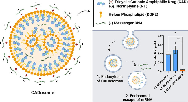
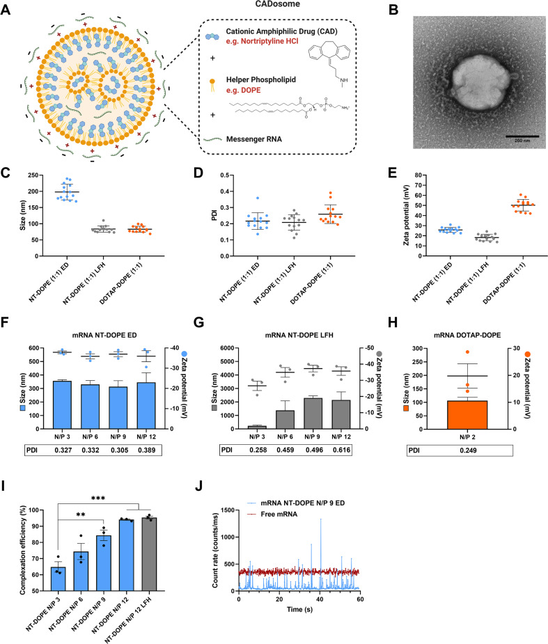
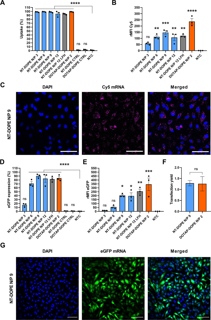
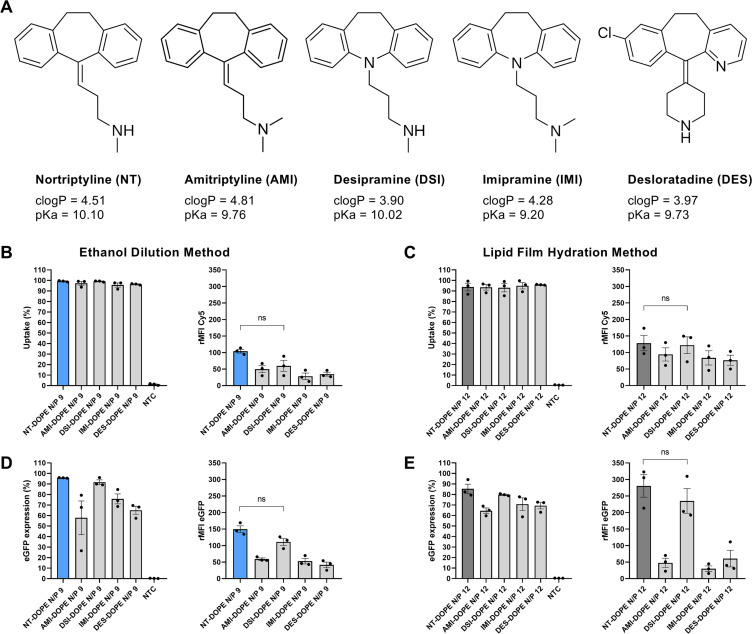
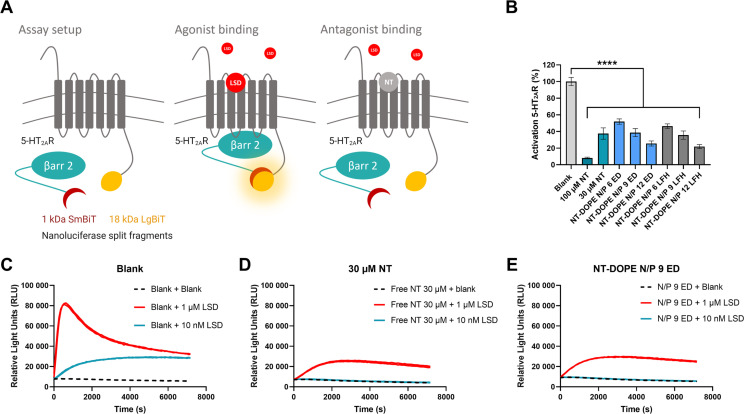
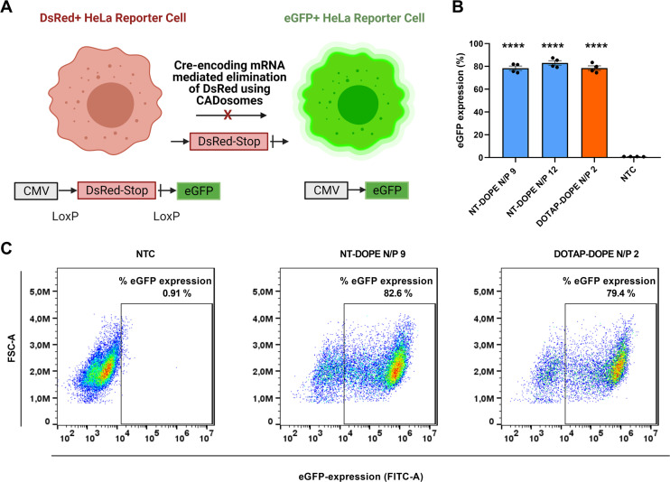
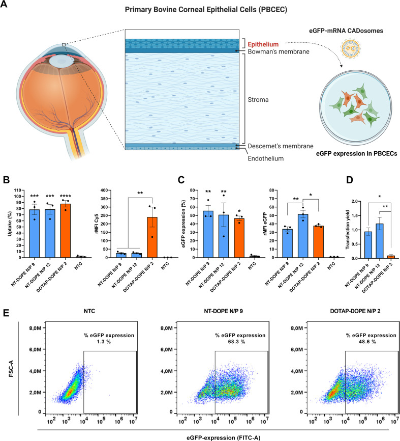
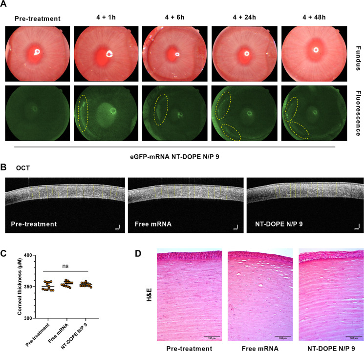
Since the recent clinical approval of siRNA-based drugs and COVID-19 mRNA vaccines, the potential of RNA therapeutics for patient healthcare has become widely accepted. Lipid nanoparticles (LNPs) are currently the most advanced nanocarriers for RNA packaging and delivery. Nevertheless, the intracellular delivery efficiency of state-of-the-art LNPs remains relatively low and safety and immunogenicity concerns with synthetic lipid components persist, altogether rationalizing the exploration of alternative LNP compositions. In addition, there is an interest in exploiting LNP technology for simultaneous encapsulation of small molecule drugs and RNA in a single nanocarrier. Here, we describe how well-known tricyclic cationic amphiphilic drugs (CADs) can be repurposed as both structural and functional components of lipid-based NPs for mRNA formulation, further referred to as CADosomes. We demonstrate that selected CADs, such as tricyclic antidepressants and antihistamines, self-assemble with the widely-used helper lipid DOPE to form cationic lipid vesicles for subsequent mRNA complexation and delivery, without the need for prior lipophilic derivatization. Selected CADosomes enabled efficient mRNA delivery in various in vitro cell models, including easy-to-transfect cancer cells (e.g. human cervical carcinoma HeLa cell line) as well as hard-to-transfect primary cells (e.g. primary bovine corneal epithelial cells), outperforming commercially available cationic liposomes and state-of-the-art LNPs. In addition, using the antidepressant nortriptyline as a model compound, we show that CADs can maintain their pharmacological activity upon CADosome incorporation. Furthermore, in vivo proof-of-concept was obtained, demonstrating CADosome-mediated mRNA delivery in the corneal epithelial cells of rabbit eyes, which could pave the way for future applications in ophthalmology. Based on our results, the co-formulation of CADs, helper lipids and mRNA into lipid-based nanocarriers is proposed as a versatile and straightforward approach for the rational development of drug combination therapies.

Keywords: Cationic amphiphilic drugs; Cellular delivery; Drug repurposing; Lipid nanoparticles; Messenger RNA; Nanomedicine; RNA therapeutics.


Conflict of interest statement

Declaration of Competing Interest The authors declare no competing financial interest.

Figures

Unlabelled Image
Graphical abstract
Fig. 1
Fig. 1
Physicochemical characterization of mRNA CADosomes containing nortriptyline (NT)-DOPE (A) Schematic representation of NT-DOPE mRNA CADosomes, produced with vesicles obtained via an ethanol dilution (ED) or lipid film hydration (LFH) method. Created with BioRender.com (B) Representative transmission electron microscopy (TEM) image of enhanced green fluorescent protein-encoding messenger RNA (eGFP-mRNA) NT-DOPE CADosomes, prepared via ED. Scale bar corresponds to 200 nm. Dynamic light scattering data (hydrodynamic size, polydispersity index (PDI) and zeta potential) of NT-DOPE and DOTAP-DOPE vesicles, (C–E) prior to (n = 14) and (F–H) after complexation with eGFP-mRNA (n = 3). (I–J) Fluorescence correlation spectroscopy (FCS) analysis of CADosome mRNA complexation efficiency with different N/P ratios. Data are represented as mean ± the standard error of the mean (SEM) for minimum three independent repeats (n ≥ 3). Statistical analysis was performed using One Way Anova with Tukey Correction (** p ≤ 0.01, *** p ≤ 0.001). (For interpretation of the references to color in this figure legend, the reader is referred to the web version of this article.)
Fig. 2
Fig. 2
Evaluating nortriptyline (NT)-DOPE CADosomes for cytosolic delivery of eGFP-encoding mRNA in a HeLa cell line. (A–B) Evaluation of cellular uptake of NT-DOPE CADosomes with different N/P ratios, loaded with Cy5-labeled mRNA, in HeLa cells as analyzed via flow cytometry and expressed as Cy5+ cells and Cy5 relative mean fluorescence intensity (rMFI Cy5; normalized to non-treated control (NTC)), respectively. (C) Representative confocal images of HeLa cells after transfection with Cy5-mRNA NT-DOPE CADosomes N/P 9, with nuclei (blue) and intracellular Cy5-mRNA (magenta). Scale bars correspond to 100 μm. (D–E) eGFP expression 24 h after transfection with NT-DOPE CADosomes N/P 3, 6, 9 and 12, DOTAP-DOPE N/P 2 and negative controls (CTRL) complexing luciferase-encoding mRNA, expressed as percentage eGFP+ HeLa cells and eGFP rMFI, respectively. (F) Transfection yield (i.e. eGFP expression normalized to intracellular mRNA dose) of NT-DOPE CADosomes N/P 9 compared to DOTAP-DOPE lipoplexes. (G) Representative confocal images of HeLa cells 24 h after transfection with eGFP-mRNA NT-DOPE CADosomes N/P 9, with nuclei (blue) and enhanced green fluorescent protein (eGFP) expression (green). Scale bars correspond to 100 μm. Data are represented as mean ± the standard error of the mean (SEM) for three independent repeats (n = 3). Statistical analysis was performed using One Way Anova with Tukey Correction (ns p > 0.05, * p ≤ 0.05, ** p ≤ 0.01, *** p ≤ 0.001, **** p ≤ 0.0001). (For interpretation of the references to color in this figure legend, the reader is referred to the web version of this article.)
Fig. 3
Fig. 3
Screening of different CADosomes for mRNA delivery in HeLa cells. (A) Molecular structure of CAD molecules which were capable of forming CADosomes in combination with DOPE (50:50 molar ratio). (B–C) Flow cytometry quantification of cellular uptake and (D–E) eGFP-mRNA expression of AMI-DOPE, DSI-DOPE, IMI-DOPE and DES-DOPE compared to NT-DOPE CADosomes, loaded with Cy5-labeled or eGFP-encoding mRNA, respectively. The latter CADosomes were prepared via ethanol dilution (B–D) and lipid film hydration method (C–E) using optimal N/P ratios. Data are represented as mean ± the standard error of the mean (SEM) for three independent repeats (n = 3). Statistical analysis was performed using One Way Anova with Tukey Correction (ns p > 0.05). (rMFI = relative mean fluorescence intensity normalized to non-treated cells (NTC)).
Fig. 4
Fig. 4
Evaluation of the pharmacological activity of nortriptyline following CADosome formulation using a Nanoluciferase Binary Technology (NanoBiT®) bioassay (A) Schematic illustration of the NanoBiT® system, with HEK293T cells stably expressing two inactive luciferase split fragments (1 kDa SmBiT and 18 kDa LgBiT), coupled to the 5-HT2AR (serotonin 2A receptor) and the cytosolic protein β-arrestin 2 (βarr2), respectively [[54], [55], [56]]. Binding of a receptor agonist, in this case LSD, results in βarr2 recruitment to the 5-HT2AR with the concomitant functional complementation of the enzyme, which can be monitored through luminescence read-out. Binding of a receptor antagonist, e.g. nortriptyline (NT), inhibits LSD-induced βarr2 recruitment and subsequent luciferase complementation. (B) Percentage 5-HT2AR activation induced by blank, free NT (30 and 100 μM) and different NT-DOPE CADosomes loaded with eGFP-mRNA, measured by calculating the normalized area under the curve (AUC) values of the receptor activation profiles. 1 μM LSD was added to all samples and luminescence was continuously monitored for 2 h. Data are represented as mean ± the standard error of the mean (SEM) for three independent repeats (n = 3). Statistical analysis was performed using One Way Anova with Tukey Correction (**** p ≤ 0.0001). (C–D–E) One representative activation profile of blank, free NT 30 μM and mRNA NT-DOPE N/P 9, respectively.
Fig. 5
Fig. 5
CADosome-mediated delivery of Cre-recombinase encoding mRNA (Cre-mRNA) in a HeLa reporter cell line. (A) Schematic illustration of HeLa reporter cells shifting from DsRed+ to eGFP+ after Cre-recombinase mediated elimination of the DsRed stop-codon following successful delivery of Cre-encoding mRNA via CADosomes. Created with BioRender.com (B) Percentage eGFP+ cells as analyzed via flow cytometry 24 h after transfection with NT-DOPE CADosomes N/P 9–12 and DOTAP-DOPE N/P 2. Data are represented as mean ± the standard error of the mean (SEM) for minimum three independent repeats (n ≥ 3). Statistical analysis was performed using One Way Anova with Tukey Correction (**** p ≤ 0.0001). (C) Representative flow cytometry dot-plots of non-treated cells (NTC) and NT-DOPE CADosomes N/P 9 or DOTAP-DOPE N/P 2 transfected HeLa reporter cells, respectively.
Fig. 6
Fig. 6
Delivery of mRNA with NT-DOPE CADosomes in primary bovine corneal epithelial cells (PBCEC). (A) Schematic illustration of the different tissue layers in the human cornea. The corneal epithelial layer was separated from corneal stroma using stainless tweezers and cultured for mRNA transfection. Created with BioRender.com (B) Cell uptake of Cy5-mRNA via NT-DOPE CADosomes N/P 9–12 was significantly lower compared to cationic DOTAP-DOPE lipoplexes after 4 h incubation. (C–D) CADosomes outperformed DOTAP-DOPE lipoplexes, reaching a ten-fold higher transfection yield for delivery of eGFP-mRNA to hard-to-transfect PBCECs, as measured 24 h after administration. (E) Representative dot-plots of non-treated cells (NTC), eGFP-mRNA NT-DOPE CADosomes N/P 9 and eGFP-mRNA DOTAP-DOPE N/P 2 lipoplexes analyzed via flow cytometry. Data are represented as mean ± the standard error of the mean (SEM) for three independent repeats (n = 3). Statistical analysis was performed using One Way Anova with Tukey Correction (* p ≤ 0.05, ** p ≤ 0.01, *** p ≤ 0.001, **** p ≤ 0.0001).
Fig. 7
Fig. 7
In vivo evaluation of eGFP-mRNA delivery and corneal toxicity of mRNA CADosomes in a rabbit model. (A) Color fundus and fluorescence images of rabbit eyes treated with eGFP-mRNA NT-DOPE N/P 9 CADosomes at a final concentration of 2 μg/mL. The marked yellow circles indicate regions with high eGFP expression at different timepoints post treatment. (B–C) 2D Optical Coherence Tomography (OCT) images visualizing the corneal thickness pre- and post-administration. No significant change could be observed (n = 12 measurements at different locations of the cornea, analyzed via ImageJ software). Scale bars correspond to 100 μm. Statistical analysis was performed using One Way Anova with Tukey Correction (ns p ≥ 0.05). (D) Histology of the corneal surface treated with free eGFP-mRNA and eGFP-mRNA CADosomes, respectively. The presented images are representative of three rabbits for each group (n = 3). (For interpretation of the references to color in this figure legend, the reader is referred to the web version of this article.)

References

    1. Kanasty R., Dorkin J.R., Vegas A., Anderson D. Delivery materials for siRNA therapeutics. Nat. Mater. 2013;12:967–977. - PubMed
    1. DeWeerdt S. RNA therapies explained. Nature. 2019;574:S2–S3.
    1. Setten R.L., Rossi J.J., Han S.P. The current state and future directions of RNAi-based therapeutics. Nat. Rev. Drug Discov. 2019;18:421–446. - PubMed
    1. Sahin U., Karikó K., Türeci Ö. mRNA-based therapeutics-developing a new class of drugs. Nat. Rev. Drug Discov. 2014;13:759–780. - PubMed
    1. Pardi N., Hogan M.J., Porter F.W., Weissman D. mRNA vaccines-a new era in vaccinology. Nat. Rev. Drug Discov. 2018;17:261–279. - PMC - PubMed

Publication types