Review
. 2022 Sep;51(3):459-481.
doi: 10.1016/j.ecl.2022.02.009. Epub 2022 Jul 8.

Lipoprotein Assessment in the twenty-first Century

Affiliations
Review

Lipoprotein Assessment in the twenty-first Century

Diego Lucero et al. Endocrinol Metab Clin North Am. 2022 Sep.

Abstract

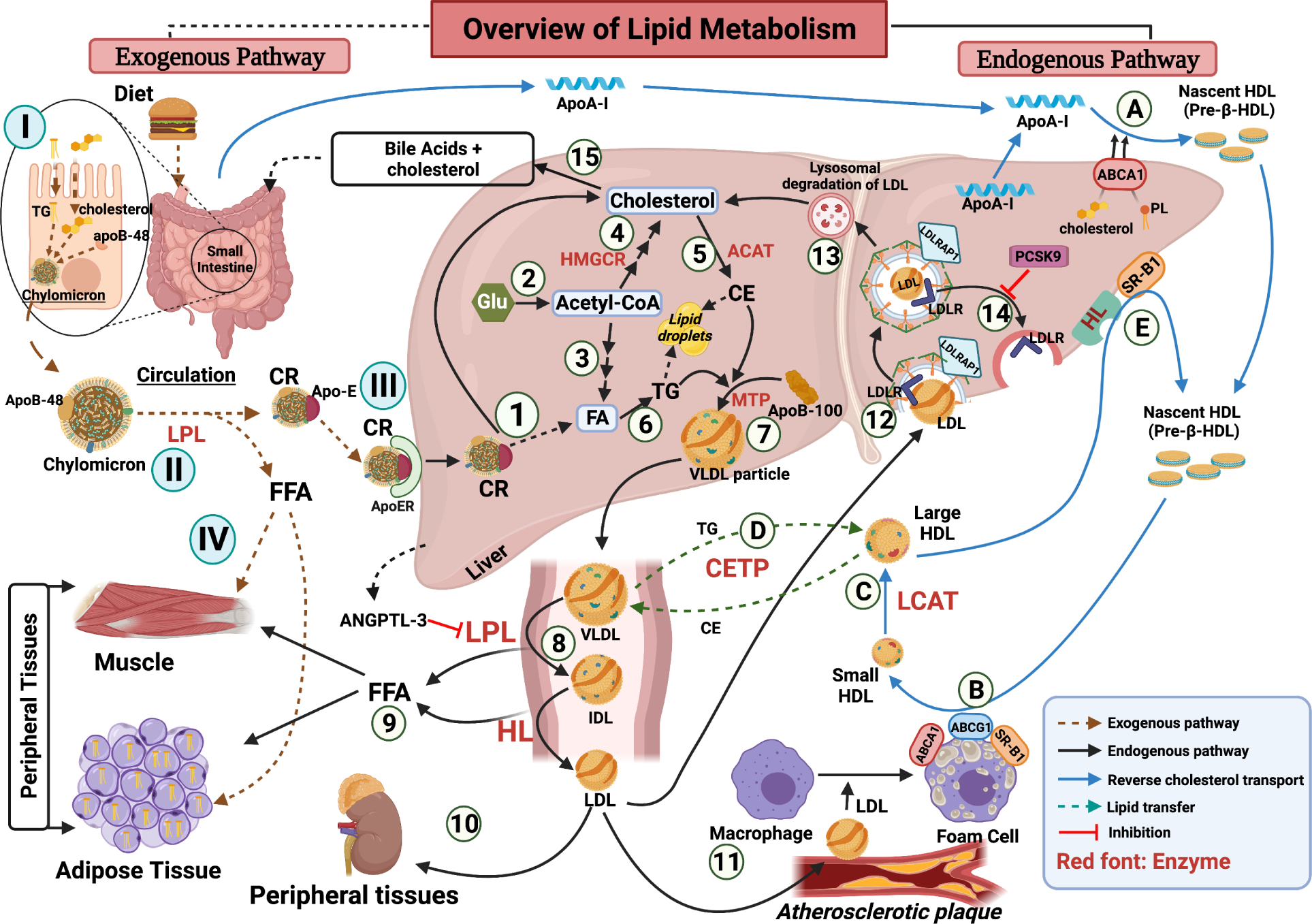
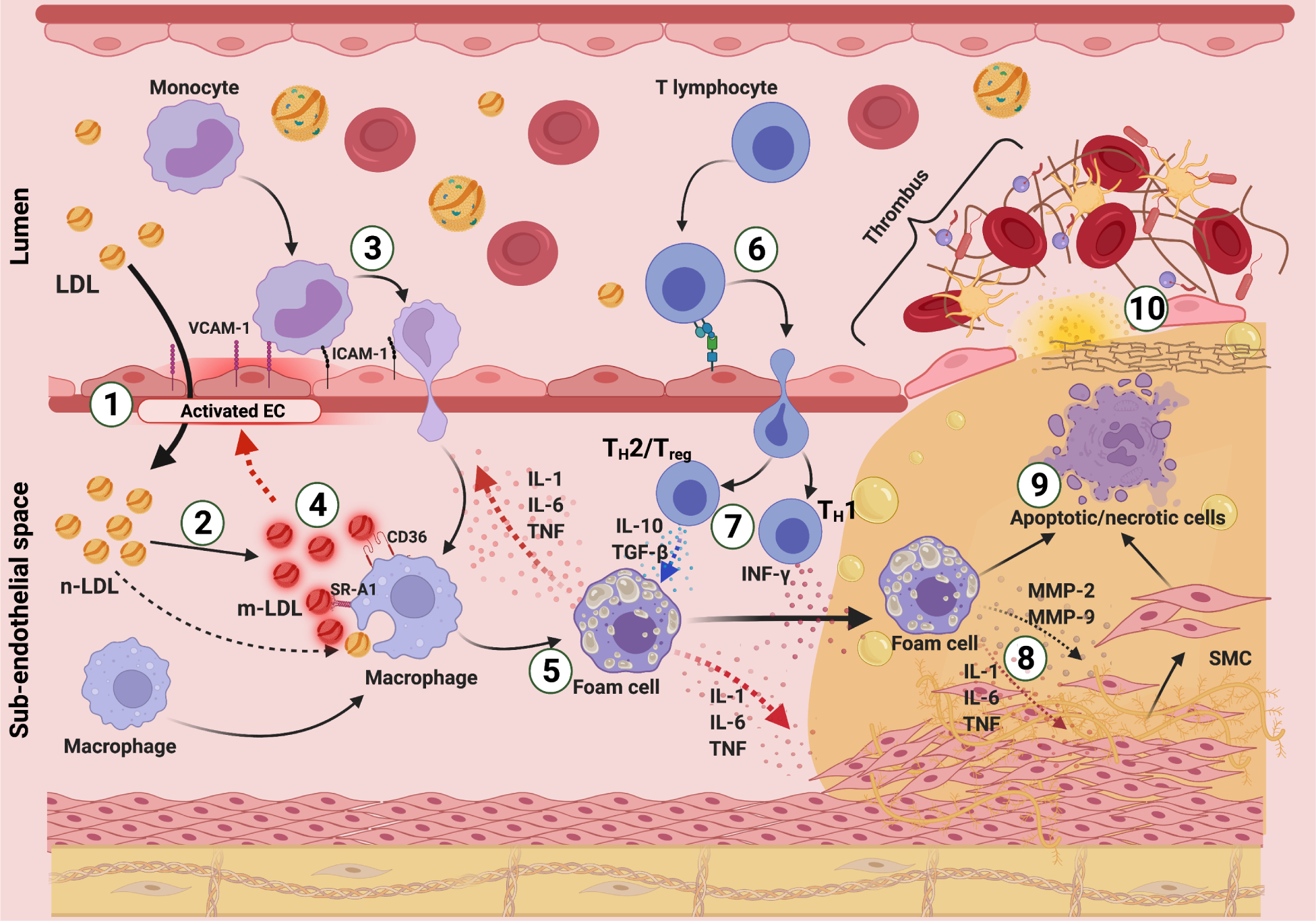
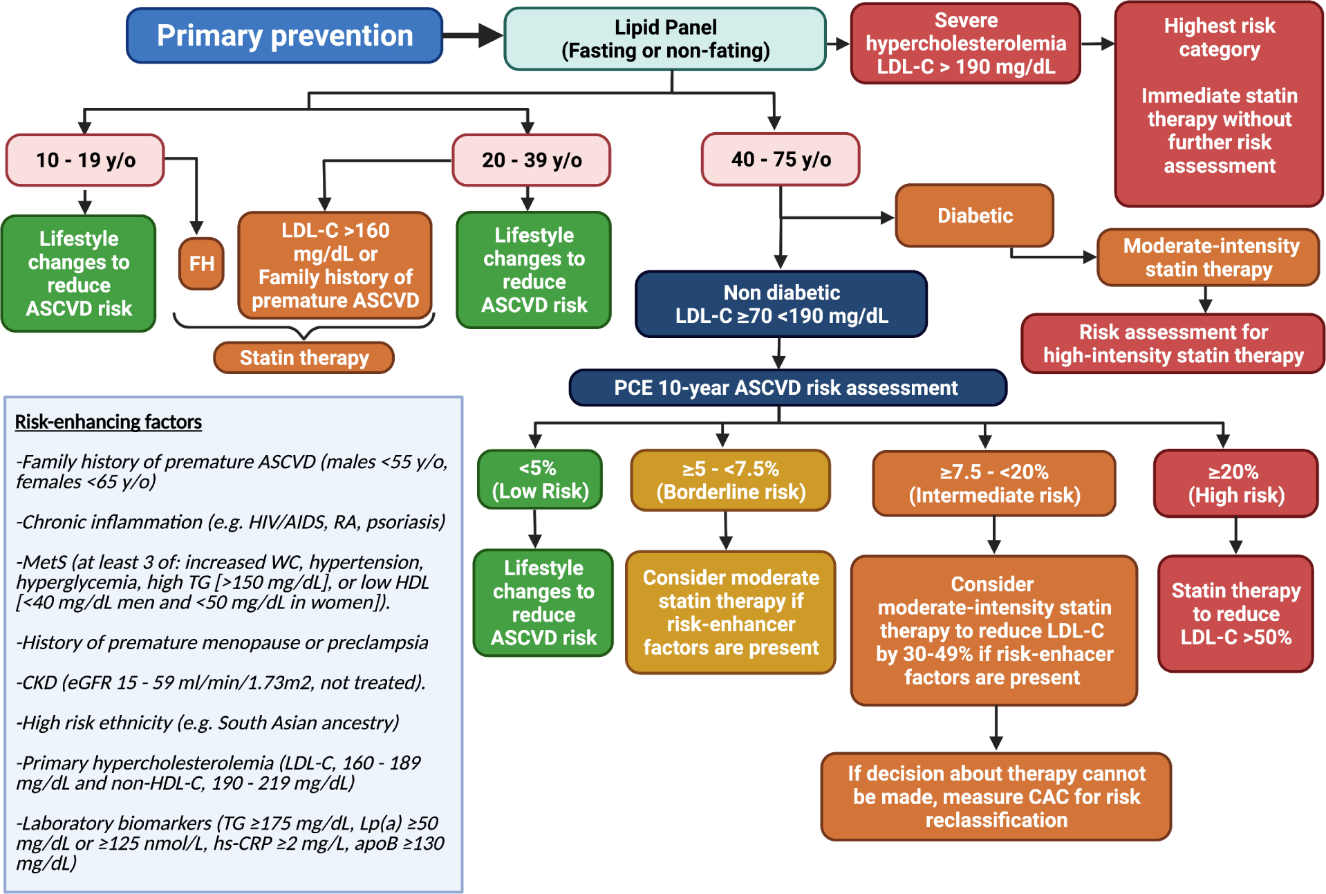
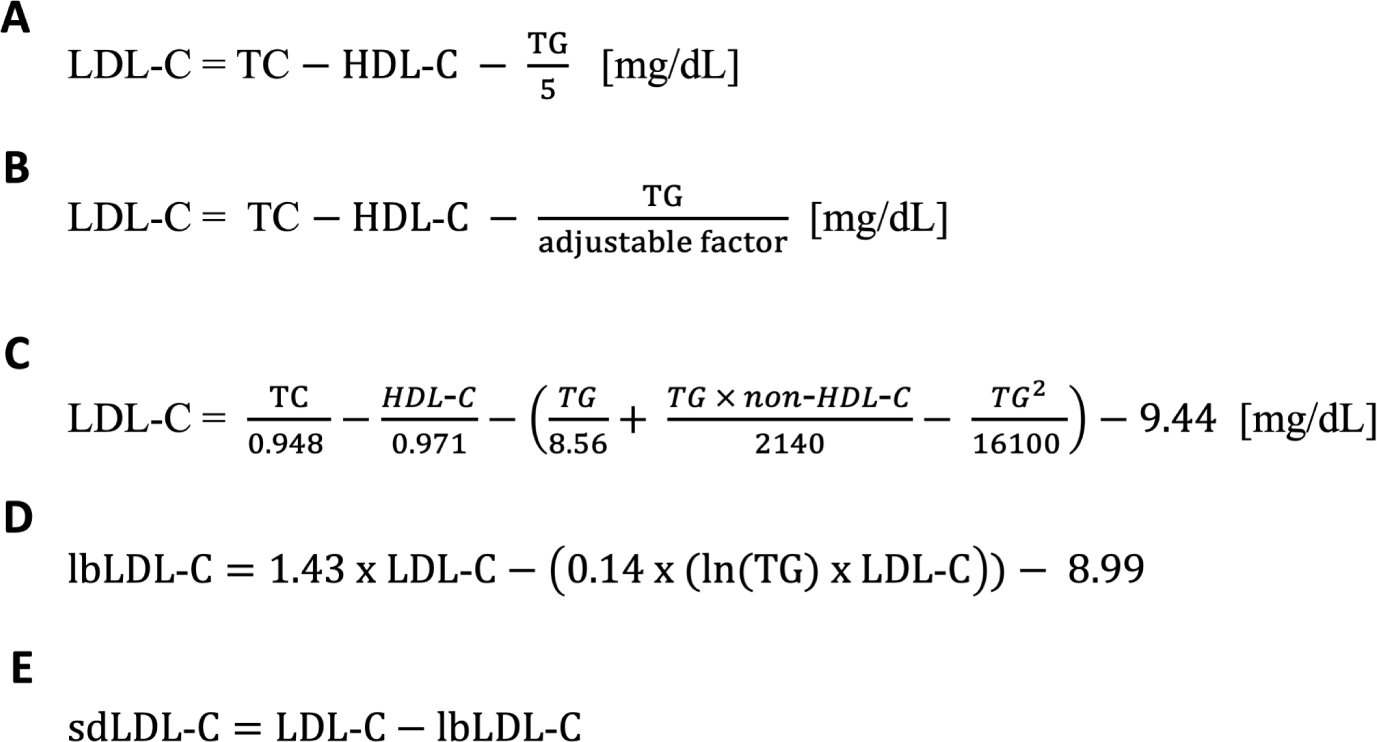
Based on decades of both basic science and epidemiologic research, there is overwhelming evidence for the causal relationship between high levels of cholesterol, especially low-density lipoprotein cholesterol and cardiovascular disease. Risk evaluation and monitoring the response to lipid-lowering therapies are heavily dependent on the accurate assessment of plasma lipoproteins in the clinical laboratory. This article provides an update of lipoprotein metabolism as it relates to atherosclerosis and how diagnostic measures of lipids and lipoproteins can serve as markers of cardiovascular risk, with a focus on recent advances in cardiovascular risk marker testing.

Keywords: Advanced lipoprotein testing; Cardiovascular risk assessment; HDL functionality; LDL-C calculation; Lipoprotein (a); Lipoprotein diagnostic assays; Non-HDL cholesterol; Recommendations for lipoprotein assessment.


Figures

Figure 1.
Figure 1.. Overview of lipoprotein metabolism.
(I – IV) Exogenous Pathway: (I) Dietary lipids are absorbed in enterocytes, packed into chylomicrons that are secreted into lymph/blood (II) In circulation, LPL lipolyze chylomicrons into chylomicron remnants (CR) and Free Fatty Acids (FFA). (III) CR, rich in apo-B48 and Apo-E, are internalized by the liver through apo-E receptor (ApoER). (IV) FFA are used or stored in peripheral tissues. (1 – 10) Endogenous Pathway: (1) CR internalized in the liver are degraded into its components. Cholesterol and fatty acids (FA) enlarge their respective hepatic pools. (2) Acetyl-CoA, coming from glucose degradation, is used for: (3) de novo synthesis FA (lipogenesis) or (4) endogenous cholesterol synthesis, regulated at 3-Hydroxy-3-Methylglutaryl-CoA Reductase (HMGCR). (5) Hepatic free cholesterol is esterified into cholesterol esters (CE) by intracellular Acyl-CoA:cholesterol acyltransferase (ACAT) and (6) FA are esterified into triglycerides (TG). TG and CE are either stored in intracellular lipid droplets or (7) added to nascent apolipoprotein B-100 (apoB-100) by microsomal triglyceride transfer protein (MTP) to form very-low density lipoprotein (VLDL), which is secreted into circulation. (8) In circulation, TG in VLDL are lipolyzed by lipoprotein lipase (LPL) forming intermediate density lipoprotein (IDL). Liver secretes angiopoietin-like 3 (ANGPTL3) that inhibits LPL in peripheral tissues. IDL is then further lipolyzed by hepatic lipase (HL) into low-density lipoprotein (LDL). (9) Released FFA are internalized by muscles or adipose tissue for oxidation or storage, respectively. LDL in circulation has three main fates: (10) internalization by peripheral tissues with high cholesterol demands (adrenal glands, testis, ovaries, etc.), (11) infiltration into the arterial wall, where it suffers modifications and is internalized by macrophages to form foams cells leading to atherosclerosis, or (12) internalization through hepatic LDL receptor (LDLR) in a clathrin-mediated endocytic process aided by the LDLR adaptor protein 1 (LDLRAP1). (13) Internalized LDL is degraded in lysosomes and its cholesterol enlarges the hepatic cholesterol pools. (14) LDLR is recycled back to the plasma membrane. This is inhibited by the proprotein convertase subtilisin/kexin type 9 (PCSK9). (15) Excess of hepatic cholesterol is secreted into the intestine as bile acids. HDL metabolism (reverse cholesterol transport) (A – E): (A) Lipid poor apolipoprotein A-I (apoA-I) is secreted by liver and small intestine. Free lipid poor apoA-I acquires cholesterol and phospholipids (PL) through hepatic ATP binding cassette (ABC) subfamily A member 1 (ABCA1) and forms nascent discoidal high-density lipoprotein (HDL). (B) Nascent HDL captures free cholesterol and phospholipids from the surface of foam cells and other peripheral cells through surface transporters (ABCA1, ABC subfamily G member 1 (ABCG1), and Scavenger Receptor class B type I (SR-BI)), forming small spherical HDL particles. (C) Lecithin-cholesterol acyltransferase (LCAT) esterifies free cholesterol in small HDL and forms large mature HDL particles. (D) Cholesteryl ester transfer protein (CETP) transfers TG from VLDL to HDL and CE from HDL to VLDL. (E) At hepatic level, triglycerides and phospholipids in large HDL are lipolyzed by HL, or CE in large HDL are taken up by hepatic SR-BI. This process generates small HDL particles that can either re-enter the HDL’s metabolic pathway or be filter and excreted through the kidney. Created with BioRender.com
Figure 2:
Figure 2:. Pathophysiology of atherosclerosis.
(1) In areas of turbulent blood flow, high shear stress induces the activation of endothelial cells (EC) leading to an increase endothelial barrier permeability. In these areas, low-density lipoproteins (LDL) infiltrate into the sub-endothelial space. (2) In the vessel wall LDL can undergo modifications, such as oxidation and aggregation (mLDL), becoming more immunogenic and pro-inflammatory. Oxidation products further stimulate endothelial activation. (3) Activated endothelium express adhesion molecules (vascular cell adhesion molecule 1 (VCAM-1) and intracellular adhesion molecules 1 (ICAM-1)) on the luminal surface. Circulating monocytes interact with adhesion molecules and transmigrate to the sub-endothelial space where they differentiate into macrophages. (4) Macrophages internalize mLDL by receptor-mediated endocytosis, involving scavenger receptors (SR-A1 and CD36), lectin-like oxidized LDL 1 (LOX-1) receptor, and LDL receptor-related protein 1 (LRP1), and native LDL by fluid-phase endocytosis. (5) Macrophages transform into cholesterol-loaded foam cells that secrete pro-inflammatory cytokines such as interleukin 1 (IL-1), IL-6, and tumor necrosis factor (TNF), which amplifies the inflammatory response and promote the proliferation of smooth muscle cells (SMC). (6) Adaptative immune cells infiltrate into the lesion area. (7) While type 1 T helper lymphocytes (TH1) secrete interferon gamma IFN-γ and exacerbate the inflammation, TH2 and Treg cells secrete anti-inflammatory cytokines (IL-10 and transforming growth factor beta (TGF-ß)) to limit inflammation and SMC proliferation. (8) Inflammation and lipid accumulation continue, and foam cells secrete metalloproteinases (MMP-2 and MMP-9) that degrade the proteoglycan matrix, favoring the migration of SMC. (9) Apoptosis and necrosis of foam cells and SMC lead to the formation of the necrotic core. (10) Plaques with large lipid accumulation and thin fibrous cap are more prone to rupture. Plaque’s content is exposed, triggering thrombosis which can occlude the artery lumen and cause downstream ischemia. Created with BioRender.com
Figure 3.
Figure 3.. Primary Prevention approach in the US 2018 Multi-Society Guidelines.
LDL-C: low-density lipoprotein cholesterol. ASCVD: Atherosclerotic Cardiovascular Disease. FH: Familial Hypercholesterolemia. PCE: Pooled Cohort Equations. HIV/AIDS: Human Immunodeficiency Virus/Acquired Immunodeficiency Syndrome. RA: Rheumatoid Arthritis. MetS: Metabolic Syndrome. WC: Waist Circumference. TG: triglycerides. HDL: High-density lipoproteins. CKD: Chronic Kidney Disease. eGFR: Estimated Glomerular Filtration Rate. Non-HDL-C: Non-HDL Cholesterol. Lp(a): Lipoprotein (a). hs-CRP: high sensitivity “C” Reactive Protein. Adapted from Grundy, S.M., et al., 2018 AHA/ACC/AACVPR/AAPA/ABC/ACPM/ADA/AGS/APhA/ASPC/NLA/PCNA Guideline on the Management of Blood Cholesterol: A Report of the American College of Cardiology/American Heart Association Task Force on Clinical Practice Guidelines. Circulation, 2019. 139(25): p. e1082-e1143.
Figure 4.
Figure 4.. Equations for the calculation of total low-density lipoprotein cholesterol (LDL-C), large buoyant LDL-C (lbLDL-C), and small dense LDL-C (sdLDL-C).
TC: total cholesterol, HDL-C: high-density lipoprotein cholesterol, TG: triglycerides, non-HDL-C: non-high-density lipoprotein cholesterol, adjustable factor: median ratio between TG and very low-density lipoprotein cholesterol categorized by TG and non-HDL-C [35].

Similar articles

Cited by

References

    1. Goldstein JL and Brown MS, A century of cholesterol and coronaries: from plaques to genes to statins. Cell, 2015. 161(1): p. 161–172. - PMC - PubMed
    1. Feingold KR, Introduction to Lipids and Lipoproteins, in Endotext, Feingold KR, et al., Editors. 2000: South Dartmouth (MA).
    1. Barale C, et al., PCSK9 Biology and Its Role in Atherothrombosis. Int J Mol Sci, 2021. 22(11). - PMC - PubMed
    1. Gennemark P, et al., An oral antisense oligonucleotide for PCSK9 inhibition. Sci Transl Med, 2021. 13(593). - PubMed
    1. Pettersen D and Fjellstrom O, Small molecule modulators of PCSK9 - A literature and patent overview. Bioorg Med Chem Lett, 2018. 28(7): p. 1155–1160. - PubMed

Publication types

MeSH terms