

Determining the existence of the foramen of Huschke in patients with temporomandibular joint disorders using cone beam computed tomography: retrospective cohort study
- PMID: 35963990
- PMCID: PMC9375943
- DOI: 10.1186/s12880-022-00850-1
Determining the existence of the foramen of Huschke in patients with temporomandibular joint disorders using cone beam computed tomography: retrospective cohort study
Abstract
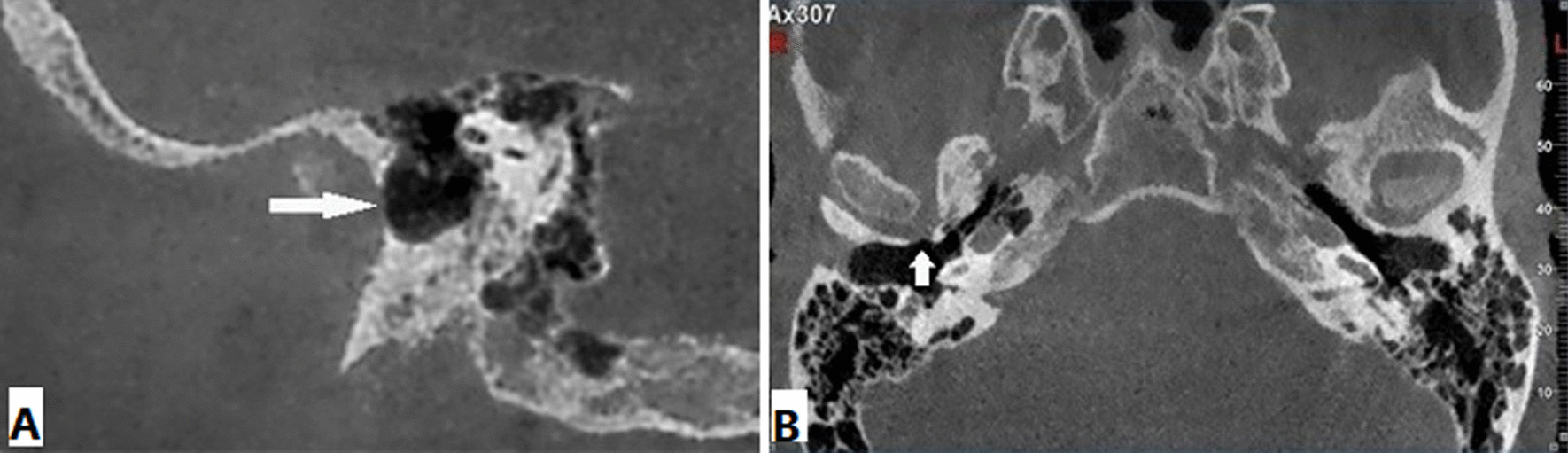
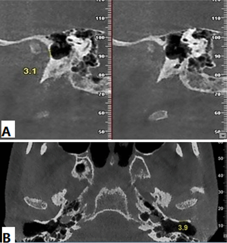
Background: Foramen of Huschke has been presented as an unusual developmental defect in anteroinferior aspect of external auditory canal. It can be associated with significant otologic complications. The purpose of this study was to determine the association between existence of foramen of Huschke and temporomandibular joint disorders in Cone Beam Computed Tomography (CBCT) images.
Methods: Of an initial sample of 465 patients, we retrospectively evaluated the CBCT images of 118 individuals with clinical signs and symptoms of temporomandibular joint disorders as case group and 256 individuals as control group. The presence, size and localization of foramen of Huschke were assessed in the axial and corrected sagittal images. The sex and age distribution were determined. Fisher's exact test, T-test and Pearson's Chi-square were applied to assess the relationship between foramen of Huschke and temporomandibular joint disorders in the case and control groups considering age and sex.
Results: The foramen of Huschke prevalence was slightly higher in patients with temporomandibular joint disorders (3.4%) than patients without temporomandibular joint disorders (0.8%). However, the difference was not statistically significant (P = 0.082). foramen of Huschke was found in five females and one male. There was no significant difference between case and control groups considering the age of patients with foramen of Huschke (P = 0.683). There was no significant difference between the case and control groups, considering the right and left ears in distribution of foramen of Huschke (P = 0.099) (P = 0.183).
Conclusions: Higher prevalence of foramen of Huschke in patients with temporomandibular joint disorders may suggest possible mechanism for temporomandibular joint disorders development that can be affected by presence of foramen of Huschke.
Keywords: Cone beam computed tomography; External auditory canal; Foramen of Huschke; Temporomandibular disorders.
© 2022. The Author(s).
Conflict of interest statement
The authors declare that they have no conflict of interest.
Figures
Similar articles
-
Prevalence of foramen Huschke: evaluation of the association between mastoid pneumatization volume and the existence of foramen Huschke using cone beam computed tomography.Eur Arch Otorhinolaryngol. 2021 Mar;278(3):791-796. doi: 10.1007/s00405-020-06296-x. Epub 2020 Aug 19. Eur Arch Otorhinolaryngol. 2021. PMID: 32813172
-
Perspective on Temporomandibular Joint Disorder: Foramen Tympanicum Defect.J Oral Rehabil. 2024 Jun;51(6):992-997. doi: 10.1111/joor.13677. Epub 2024 Mar 3. J Oral Rehabil. 2024. PMID: 38433411
-
Foramen tympanicum or foramen of Huschke: anatomical cone beam CT study.Dentomaxillofac Radiol. 2012 May;41(4):294-7. doi: 10.1259/dmfr/62359484. Dentomaxillofac Radiol. 2012. PMID: 22517996 Free PMC article.
-
Persistent foramen of Huschke: Clinical manifestations and complications, systematic review.J Stomatol Oral Maxillofac Surg. 2023 Dec;124(6):101455. doi: 10.1016/j.jormas.2023.101455. Epub 2023 Mar 24. J Stomatol Oral Maxillofac Surg. 2023. PMID: 36965816
-
Developmental defects of the tympanic plate: case reports and review of the literature.J Oral Maxillofac Surg. 1989 Dec;47(12):1336-40. doi: 10.1016/0278-2391(89)90738-6. J Oral Maxillofac Surg. 1989. PMID: 2685214 Review.
Cited by
-
The relation between persistent foramen tympanicum and degenerative bone alterations in temporomandibular joint region.Oral Radiol. 2024 Jul;40(3):445-453. doi: 10.1007/s11282-024-00749-3. Epub 2024 Apr 8. Oral Radiol. 2024. PMID: 38587690
-
Malignant myoepithelioma of the external auditory canal - a rare case report with literature review and clinical importance of foramen of Huschke.World J Surg Oncol. 2024 Jan 24;22(1):28. doi: 10.1186/s12957-024-03317-5. World J Surg Oncol. 2024. PMID: 38268020 Free PMC article. Review.
-
Association of Anatomical Features of the Petrotympanic Fissure and Presence of Foramen of Huschke With Temporomandibular Disorders.J Oral Rehabil. 2025 May;52(5):597-603. doi: 10.1111/joor.13923. Epub 2025 Jan 9. J Oral Rehabil. 2025. PMID: 39789824 Free PMC article.
References
-
- Duman ŞB, Yeşiltepe S, Bayrakdar İŞ, Yaşa Y-s. Retrospective comparison of cleft lip/palate patients and normal controls: cone beam computed tomograph imaging of foramen Husckhe morphology. Eur J Anat. 2020;24:31–5.
-
- Wang R-G, Bingham B, Hawke M, Kwok P, Li J. Persistence of the foramen of Huschke in the adult: an osteological study. J Otolaryngol. 1991;20:251. - PubMed
-
- Hawke M, Kwok P, Mehta M, Wang R-G. Bilateral spontaneous temporomandibular joint herniation into the external auditory canal. J Otolaryngol. 1987;16:387–9. - PubMed
Publication types
MeSH terms
LinkOut - more resources
Full Text Sources
Medical
