. 2022 Aug 13;13(1):413.
doi: 10.1186/s13287-022-03105-6.

Intra-amniotic transplantation of brain-derived neurotrophic factor-modified mesenchymal stem cells treatment for rat fetuses with spina bifida aperta

Affiliations

Intra-amniotic transplantation of brain-derived neurotrophic factor-modified mesenchymal stem cells treatment for rat fetuses with spina bifida aperta

Wei Ma et al. Stem Cell Res Ther. .

Abstract

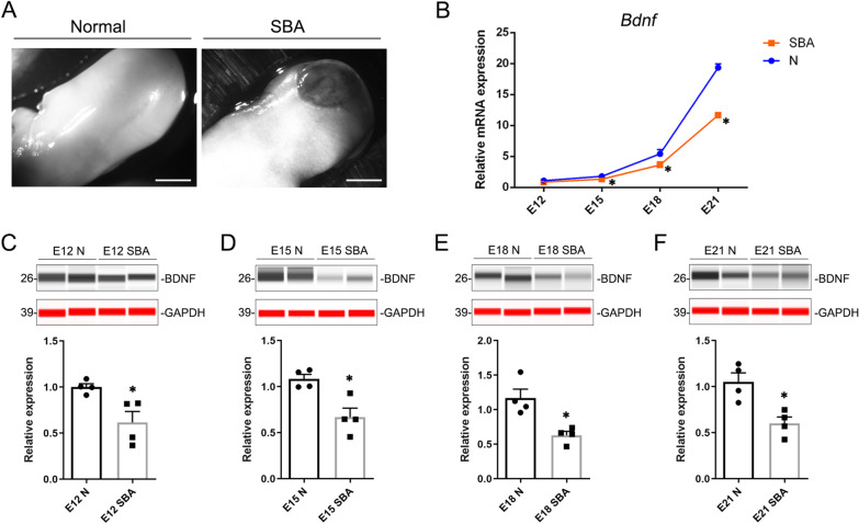
Background: Spina bifida aperta (SBA) is a relatively common clinical type of neural tube defect. Although prenatal fetal surgery has been proven to be an effective treatment for SBA, the recovery of neurological function remains unsatisfactory due to neuron deficiencies. Our previous results demonstrated that intra-amniotic transplanted bone marrow mesenchymal stem cells (BMSCs) could preserve neural function through lesion-specific engraftment and regeneration. To further optimize the role of BMSCs and improve the environment of defective spinal cords so as to make it more conducive to nerve repair, the intra-amniotic transplanted BMSCs were modified with brain-derived neurotrophic factor (BDNF-BMSCs), and the therapeutic potential of BDNF-BMSCs was verified in this study.

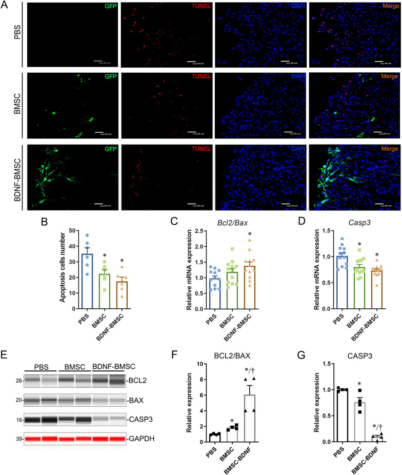
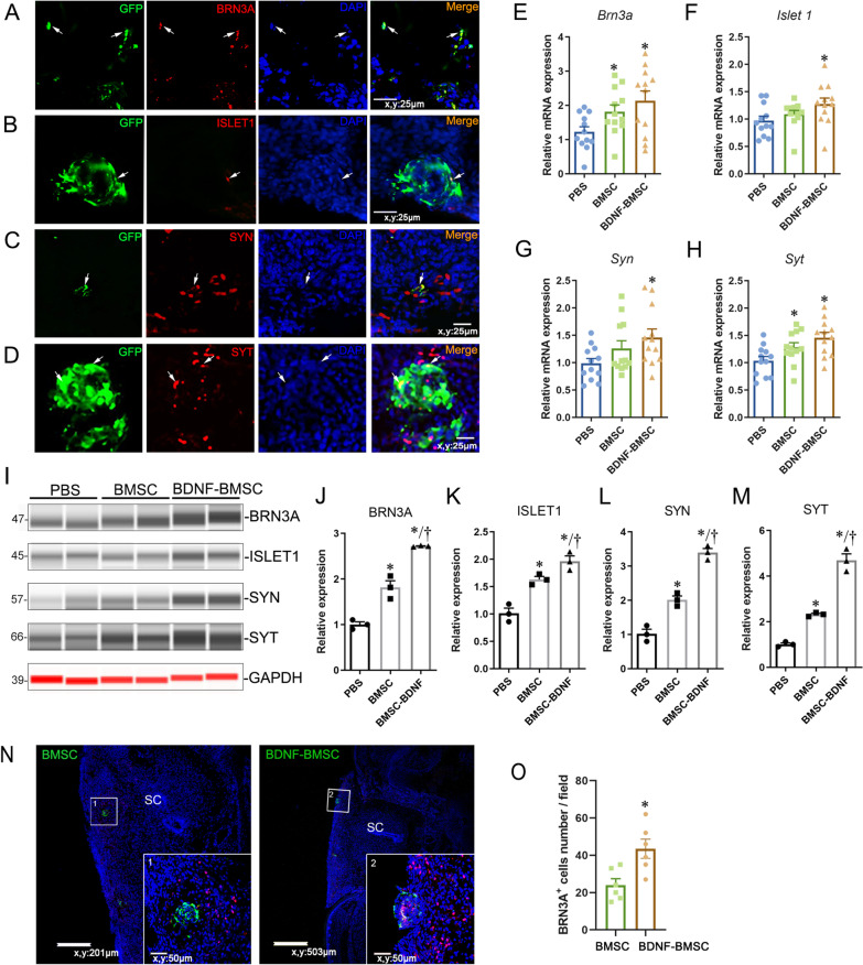
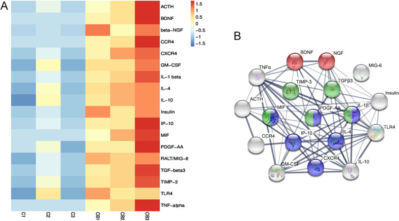
Methods: BMSCs were modified by adenovirus encoding a green fluorescent protein and brain-derived neurotrophic factor (Ad-GFP-BDNF) in vitro and then transplanted into the amniotic cavity of rat fetuses with spina bifida aperta which were induced by all-trans-retinoic acid on embryonic day 15. Immunofluorescence, western blot and real-time quantitative PCR were used to detect the expression of different neuron markers and apoptosis-related genes in the defective spinal cords. Lesion areas of the rat fetuses with spina bifida aperta were measured on embryonic day 20. The microenvironment changes after intra-amniotic BDNF-BMSCs transplantation were investigated by a protein array with 90 cytokines.

Results: We found that BDNF-BMSCs sustained the characteristic of directional migration, engrafted at the SBA lesion area, increased the expression of BDNF in the defective spinal cords, alleviated the apoptosis of spinal cord cells, differentiated into neurons and skin-like cells, reduced the area of skin lesions, and improved the amniotic fluid microenvironment. Moreover, the BDNF-modified BMSCs showed a better effect than pure BMSCs on the inhibition of apoptosis and promotion of neural differentiation.

Conclusion: These findings collectively indicate that intra-amniotic transplanted BDNF-BMSCs have an advantage of promoting the recovery of defective neural tissue of SBA fetuses.

Keywords: Brain-derived neurotrophic factor; Intra-amniotic transplantation; Mesenchymal stem cells derived from bone marrow; Prenatal treatment; Spina bifida aperta.


Conflict of interest statement

The authors indicated no potential conflicts of interest.

Figures

Fig. 1
Fig. 1
Expression pattern of BDNF in the spinal cords of normal and SBA rat fetuses. A. A typical back photograph from a normal fetus and an SBA fetus induced by atRA. B. Expression-level analysis of BDNF mRNA in normal and SBA spinal cords on E12, E15, E18, and E21. CF. Immunodetection of BDNF protein in the spinal cords of normal and SBA fetuses at E12, E15, E18, and E21 by simple western shows a significant reduction in BDNF protein in the SBA spinal cords. Gray–white stripes were detected by the HRP channel; red stripes were detected by the NIR channel. The stripes and quantification of relative protein levels were determined from peak areas detected by a size-based capillary electrophoresis instrument. Abbreviations: N: normal fetal rat spinal cord, SBA: spina bifida aperta, E: embryonic days, *p < 0.05
Fig. 2
Fig. 2
Intra-amniotic transplanted BDNF-BMSCs implanted into defective spinal cord and skin. A. Typical images observed under a stereoscopic fluorescence microscope; the white box is magnified, and the scale: 2.5 mm; the yellow dotted line indicates the location of the rightmost frozen section which shows that the transplanted bone marrow mesenchymal stem cells (BMSCs) engrafted into the defective spinal cord and the edge of the broken skin, scale: 100 μm. B: Quantitative analysis of the relative mRNA expression of BDNF in SBA spinal cords of PBS-injected, BMSC-injected and BDNF-BMSC-injected fetuses (n = 12 per group). C: The protein expression of BDNF and GAPDH in the defective spinal cord was detected by the simple western system in PBS-, BMSC-, and BDNF-BMSC-injected groups. D. Quantification of relative protein levels of BDNF in different transplantation groups from Figure C. *Compared to the PBS-injected group, p < 0.05; †Compared to the BMSC-injected group, p < 0.05. E. Representative images of E20 rat fetus with SBA after PBS injection, BMSC transplantation, and BDNF-BMSC transplantation taken by fluorescent stereomicroscopy; the transplanted cells were labeled by GFP. The white dots indicate the edge of the defective skin lesions. F. Quantitative analysis of the area of skin lesions in the BDNF-BMSC-injected, BMSC-injected, and PBS-injected groups (n = 23 per group). *Compare to the PBS-injected group, p < 0.05
Fig. 3
Fig. 3
Engrafted BDNF-BMSC significantly reduced the apoptosis in defective spinal cords. A. Typical images of double staining of TUNEL and GFP antibody in the spinal dorsal horn were observed in the BDNF-BMSC-injected group, the BMSC-injected group, and the PBS-injected group under a fluorescence microscope. Scale bar: 30 μm. B. Quantification of apoptotic cells was performed under 40 × field (n = 6/group). The apoptotic cells in the dorsal spinal cord of the BDNF-BMSC-injected group and BMSC-injected group were significantly decreased compared to the PBS injection group. CD. Quantitative analysis of the relative mRNA expression of BCL2, BAX, and CASP3 in the spinal cords of PBS-injected, BMSC-injected, and BDNF-BMSC-injected fetuses (n = 12/group). *The difference was significant compared to the PBS group (p < 0.05). E. Simple western detection of BCL2, BAX, and CASP3 protein expression in the spinal cords from SBA fetuses after intra-amniotic injection of PBS, BMSCs, and BDNF-BMSCs. Gray–white stripes were detected by the HRP channel; red stripes were detected by the NIR channel. FG. Quantification of the relative protein levels determined from the special peak area of BCL2, BAX, and CASP3 shown in E. *Significant difference compared to the PBS-injected group, †Significant difference compared to the BMSC-transplanted group, p < 0.05
Fig. 4
Fig. 4
Engrafted BDNF-BMSCs expressed neuron-related specific markers in the spinal cord of SBA. AD. Double fluorescent staining of BRN3A, ISLET1, SYT, and SYN with GFP in the sections of defective spinal cords with BDNF-BMSCs engraftment. Typical double-positive cells are labeled with arrows. EH. The relative mRNA expression of BRN3A, ISLET1, SYN, and SYT in the spinal cords of BDNF-BMSC-, BMSC-, and PBS-injected groups was quantitatively analyzed by RT-qPCR (n = 12/group). I. Simple western system detection of BRN3A, ISLET1, SYN, and SYT protein in the spinal cords from SBA fetuses after intra-amniotic injection of PBS, BMSCs, and BDNF-BMSCs. Gray–white stripes were detected by the HRP channel, and red stripes were detected the by NIR channel. JM. Quantification of relative protein levels determined from the special peak area of BRN3A, ISLET1, SYN, and SYT shown in I. N. Representative images of GFP and BRN3A double staining in defective spinal cords with BDNF-BMSCs and pure BMSC engraftment. The images in the small white box are enlarged in the lower right corner. SC: spinal cord. O. The BRN3A+ cells around the engrafted BMSCs-BDNF or BMSCs in defective spinal cords were counted in 40 × field (n = 6, p < 0.05). *Significant difference compared to the PBS-injected group, †Significant difference compared to the BMSC-injected group, p < 0.05
Fig. 5
Fig. 5
Engrafted BDNF-BMSCs expressed the marker protein K19 of epidermal cells. AB. Double immunofluorescence staining of GFP and K19 in frozen sections of rat fetuses with intra-amniotic transplanted BMSC and BDNF-BMSC engrafted. The white box was enlarged on the right side with three color channels. The representative double-positive cells are marked by arrows. Scale bar: 25 μm
Fig. 6
Fig. 6
Changes in the amniotic fluid microenvironment after transplantation of BDNF-BMSCs. A. Protein array analysis of amniotic fluid with BMSC transplantation (n = 3, samples: C1, C2, C3) and BDNF-BMSCs transplantation (n = 3, samples: CB1, CB2, CB3). Upregulated proteins (fold change > 3) with p < 0.05 are shown in the cluster analysis. B. Interaction network of differentially expressed proteins generated by STRING database. The colors of the inside nodes indicate that the proteins come from different biological processes, including negative regulation of neuronal apoptosis (red, GO: 0,043,524), positive regulation of cell migration (blue, GO: 0,030,335), and wound healing (green, GO: 0,042,060), while the proteins that were not involved in the above-mentioned three processes are marked in gray. The thickness of the lines connecting the nodes indicates the strength of data support

Similar articles

Cited by

References

    1. Brea CM, Munakomi S. Spina Bifida. Treasure Island (FL): StatPearls; 2022. - PubMed
    1. Adzick NS, Sutton LN, Crombleholme TM, et al. Successful fetal surgery for spina bifida. Lancet. 1998;352:1675–1676. doi: 10.1016/S0140-6736(98)00070-1. - DOI - PubMed
    1. Tulipan N, Hernanz-Schulman M, Bruner JP. Reduced hindbrain herniation after intrauterine myelomeningocele repair: a report of four cases. Pediatr Neurosurg. 1998;29:274–278. doi: 10.1159/000028735. - DOI - PubMed
    1. Sutton LN, Adzick NS, Bilaniuk LT, et al. Improvement in hindbrain herniation demonstrated by serial fetal magnetic resonance imaging following fetal surgery for myelomeningocele. JAMA. 1999;282:1826–1831. doi: 10.1001/jama.282.19.1826. - DOI - PubMed
    1. Dewan MC, Wellons JC. Fetal surgery for spina bifida. J Neurosurg Pediatr. 2019;24:105–114. doi: 10.3171/2019.4.PEDS18383. - DOI - PubMed

Publication types

Substances

LinkOut - more resources