Domain-specific connectivity drives the organization of object knowledge in the brain
- PMID: 35964974
- PMCID: PMC11498098
- DOI: 10.1016/B978-0-12-823493-8.00028-6
Domain-specific connectivity drives the organization of object knowledge in the brain
Abstract
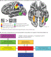
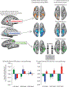
The goal of this chapter is to review neuropsychological and functional MRI findings that inform a theory of the causes of functional specialization for semantic categories within occipito-temporal cortex-the ventral visual processing pathway. The occipito-temporal pathway supports visual object processing and recognition. The theoretical framework that drives this review considers visual object recognition through the lens of how "downstream" systems interact with the outputs of visual recognition processes. Those downstream processes include conceptual interpretation, grasping and object use, navigating and orienting in an environment, physical reasoning about the world, and inferring future actions and the inner mental states of agents. The core argument of this chapter is that innately constrained connectivity between occipito-temporal areas and other regions of the brain is the basis for the emergence of neural specificity for a limited number of semantic domains in the brain.
Keywords: Category-specificity; Concepts; Domain-specificity; Neural specificity; Objects; Occipito-temporal cortex; Ventral visual pathway.
Copyright © 2022 Elsevier B.V. All rights reserved.
Figures


Similar articles
-
High-Level Representations in Human Occipito-Temporal Cortex Are Indexed by Distal Connectivity.J Neurosci. 2021 May 26;41(21):4678-4685. doi: 10.1523/JNEUROSCI.2857-20.2021. Epub 2021 Apr 13. J Neurosci. 2021. PMID: 33849949 Free PMC article.
-
The relative contributions of visual and semantic information in the neural representation of object categories.Brain Behav. 2019 Oct;9(10):e01373. doi: 10.1002/brb3.1373. Epub 2019 Sep 27. Brain Behav. 2019. PMID: 31560175 Free PMC article.
-
Action Categories in Lateral Occipitotemporal Cortex Are Organized Along Sociality and Transitivity.J Neurosci. 2017 Jan 18;37(3):562-575. doi: 10.1523/JNEUROSCI.1717-16.2016. J Neurosci. 2017. PMID: 28100739 Free PMC article.
-
Visual objects and their colors.Handb Clin Neurol. 2022;187:179-189. doi: 10.1016/B978-0-12-823493-8.00022-5. Handb Clin Neurol. 2022. PMID: 35964971 Review.
-
Division of labor between lateral and ventral extrastriate representations of faces, bodies, and objects.J Cogn Neurosci. 2011 Dec;23(12):4122-37. doi: 10.1162/jocn_a_00091. Epub 2011 Jul 7. J Cogn Neurosci. 2011. PMID: 21736460 Review.
Cited by
-
Graspable foods and tools elicit similar responses in visual cortex.bioRxiv [Preprint]. 2024 Feb 22:2024.02.20.581258. doi: 10.1101/2024.02.20.581258. bioRxiv. 2024. Update in: Cereb Cortex. 2024 Sep 3;34(9):bhae383. doi: 10.1093/cercor/bhae383. PMID: 38529495 Free PMC article. Updated. Preprint.
-
Reciprocal interactions among parietal and occipito-temporal representations support everyday object-directed actions.Neuropsychologia. 2024 Jun 6;198:108841. doi: 10.1016/j.neuropsychologia.2024.108841. Epub 2024 Feb 29. Neuropsychologia. 2024. PMID: 38430962 Free PMC article.
-
Individual variation in the functional lateralization of human ventral temporal cortex: Local competition and long-range coupling.Imaging Neurosci (Camb). 2025 Mar 3;3:imag_a_00488. doi: 10.1162/imag_a_00488. eCollection 2025 Mar 1. Imaging Neurosci (Camb). 2025. PMID: 40078535 Free PMC article.
-
Neurobiologically informed graph theory analysis of the language system.Netw Neurosci. 2025 Apr 30;9(2):504-521. doi: 10.1162/netn_a_00443. eCollection 2025. Netw Neurosci. 2025. PMID: 40487361 Free PMC article.
-
Individual variation in the functional lateralization of human ventral temporal cortex: Local competition and long-range coupling.bioRxiv [Preprint]. 2025 Jan 7:2024.10.15.618268. doi: 10.1101/2024.10.15.618268. bioRxiv. 2025. Update in: Imaging Neurosci (Camb). 2025 Mar 03;3:imag_a_00488. doi: 10.1162/imag_a_00488. PMID: 39464049 Free PMC article. Updated. Preprint.
References
-
- Allison T, McCarthy G, Nobre A, Puce A, & Belger A (1994). Human Extrastriate Visual Cortex and the Perception of Faces, Words, Numbers, and Colors. Cerebral cortex 4, 544–554. - PubMed
Publication types
MeSH terms
Grants and funding
LinkOut - more resources
Full Text Sources

