Review
. 2022:187:381-403.
doi: 10.1016/B978-0-12-823493-8.00017-1.

Amygdala connectivity and implications for social cognition and disorders

Affiliations
Review

Amygdala connectivity and implications for social cognition and disorders

Olivia C Meisner et al. Handb Clin Neurol. 2022.

Abstract

The amygdala is a hub of subcortical region that is crucial in a wide array of affective and motivation-related behaviors. While early research contributed significantly to our understanding of this region's extensive connections to other subcortical and cortical regions, recent methodological advances have enabled researchers to better understand the details of these circuits and their behavioral contributions. Much of this work has focused specifically on investigating the role of amygdala circuits in social cognition. In this chapter, we review both long-standing knowledge and novel research on the amygdala's structure, function, and involvement in social cognition. We focus specifically on the amygdala's circuits with the medial prefrontal cortex, the orbitofrontal cortex, and the hippocampus, as these regions share extensive anatomic and functional connections with the amygdala. Furthermore, we discuss how dysfunction in the amygdala may contribute to social deficits in clinical disorders including autism spectrum disorder, social anxiety disorder, and Williams syndrome. We conclude that social functions mediated by the amygdala are orchestrated through multiple intricate interactions between the amygdala and its interconnected brain regions, endorsing the importance of understanding the amygdala from network perspectives.

Keywords: Amygdala; Anatomic connectivity; Functional connectivity; Network; Nonhuman primates; Social behavior; Social dysfunction.


Conflict of interest statement

Competing financial interests The authors declare no competing financial interests.

Figures
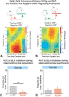
Figure 1.
Figure 1.. ACC-BLA Circuits in Social Cognition.
A–B: Dal Monte et al., 2020. A: Differences in BLA spike to ACCg LFP coherence between positive and negative ORP over time. BLA spikes and ACCg LFPs showed increased coherence in the beta frequency range for positive ORP and decreased coherence in the beta frequency band for negative ORP. B: Differences in ACCg spike to BLA LFP coherence between positive and negative ORP over time. ACCg spikes and BLA LFPs showed increased coherence in the gamma frequency range for positive ORP and decreased coherence in the gamma frequency range for negative ORP. C–D: Allsop et al., 2018. ACC→ BLA inhibition during observational fear acquisition. C: When ACC→ BLA neurons were inhibited with halorhodopsin (NpHR) during observational acquisition, NpHR mice showed significantly decreased freezing to the cue compared to control mice (eYFP) on test day. D: When ACC→ BLA neurons were inhibited with NpHR during observational fear expression, there was no significant difference between freezing behavior in control (eYFP) and NpHR mice on test day.
Figure 2.
Figure 2.. OFC-Amygdala Circuits in Social Cognition.
A–B: Munuera et al., 2018. To determine if neural representations of hierarchical rank and rewards associated with fractal images were encoded by overlapping neuronal populations, the researchers trained a linear decoder to distinguish between fractal trials with either a large reward or no reward. The decoder was then tested on held-out trials of the same type and trials in which monkeys viewed two images of monkey faces. A: Depicts the average decoding performance for Amygdala, OFC, and ACC for distinguishing between trials in which hierarchical rank of the two monkey face images presented differed. Decoding performance on monkey face trials was greater in the amygdala than in the OFC or ACC. B: Depicts the average decoding performance for Amygdala, OFC, and ACC for classifying fractal images. Decoding performance on held-out fractal trials was high for all three regions. C–D: Sato et al., 2020 C: Left amygdala to right OFC functional connectivity was positively correlated with Multidimensional Scale of Perceived Social Support (MSPSS) scores. D: Bilateral amygdala fractional amplitude of low-frequency fluctuations (fALFF) values were negatively correlated with MSPSS scores.
Figure 3.
Figure 3.. Hippocampus-Amygdala Circuits in Social Cognition.
A–C: Felix-Ortiz & Tye, 2014 A: Inactivation of basolateral amygdala (BLA)→ventral hippocampus (vHPC) neurons with halorhodopsin (NpHR) increased social interactions in the resident intruder task in comparison to control (eYFP) mice. B: Activation of BLA→vHPC neurons with channelrhodopsin-2 (ChR2) decreased social interactions in the resident intruder task in comparison to control (eYFP) mice. C: Activation of BLA→vHPC neurons with ChR2 also increased self-grooming behaviors in comparison to eYFP control mice. D–H: Okyuama et al., 2016. Comparisons of social discrimination scores when investigated brain regions were optogenetically inhibited (laser ON) versus when they were not (laser OFF). D: Inhibition of vHPC neurons significantly decreased social discrimination scores. E: Inhibition of dorsal hippocampus (dHPC) neurons did not affect social discrimination scores. F: Inhibition of vHPC→Nucleus Accumbens (NAcc) projections significantly decreased social discrimination scores. G: Inhibition of vHPC→Olfactory Bulb (OB) projections did not affect social discrimination scores. H: Inhibition of vHPC→BLA projections did not affect social discrimination scores.
Figure 4:
Figure 4:. Amygdala-cortical circuit in animal model of ASD.
A-D: Huang et al., 2016. A: Quantification of increased branching of mPFC axon terminals on the BLA in pten+/− mice (vehicle), and this phenotype is not reversed by inhibiting S6K1 in adults (PF-4708671) B: Quantification of increase in synaptic boutons on mPFC axon terminals in the BLA in pten+/− mice (vehicle), and this phenotype is not reversed by inhibiting S6K1 in adults (PF-4708671) C: Details of chemogenetic system used to reduce activity in the mPFC projections to the BLA D: Reducing activity of the mPFC-BLA projections rescues behavioral deficits in a three-chamber sociability task, pten+/− mice show no preference (pten+/−) for social stimuli over non-social stimuli, and this phenotype is rescued on inhibiting activity (hM4Di).
Figure 5:
Figure 5:. Amygdala activation in Social Anxiety Disorder and Williams Syndrome.
A–B: Cremers et al., 2015. A: Self-stress reported on 11-point Likert-scale by individuals with social anxiety and controls during: baseline, when instructed they would be giving a public speech (anticipation) and when they were informed that no speech would have to be given (recovery) B: Amygdala-cortical connectivity during baseline, anticipation and recovery periods showing transient decrease in negative functional connectivity in individuals with social anxiety C: Jones et al., 2000. Individuals with Williams syndrome (WMS) are more likely to approach unfamiliar individuals than age-matched normal controls (NC (CA)) and approximately mental age-matched normal controls (NC (approx-MA)). D: Haas et al., 2009. Amygdala activation to happy (left) and fearful (right) faces in William syndrome (WS) and typically developing controls (TD)

Similar articles

Cited by

References

    1. Abivardi A & Bach DR (2017). Deconstructing white matter connectivity of human amygdala nuclei with thalamus and cortex subdivisions in vivo. Human Brain Mapping 38: 3927–3940. - PMC - PubMed
    1. Adolphs R, Tranel D & Damasio AR (1998). The human amygdala in social judgment. Nature 393: 470–474. - PubMed
    1. Adolphs R, Tranel D, Damasio H, et al. (1994). Impaired recognition of emotion in facial expressions following bilateral damage to the human amygdala. Nature 372: 669–672. - PubMed
    1. Allison T, Puce A & McCarthy G (2000). Social perception from visual cues: Role of the STS region. Trends in Cognitive Sciences. - PubMed
    1. Allsop SA, Wichmann R, Mills F, et al. (2018). Corticoamygdala Transfer of Socially Derived Information Gates Observational Learning. Cell 173: 1329–1342 e1318. - PMC - PubMed