Review
. 2022:188:393-408.
doi: 10.1016/B978-0-323-91534-2.00012-6.

The phrenic neuromuscular system

Affiliations
Review

The phrenic neuromuscular system

David D Fuller et al. Handb Clin Neurol. 2022.

Abstract

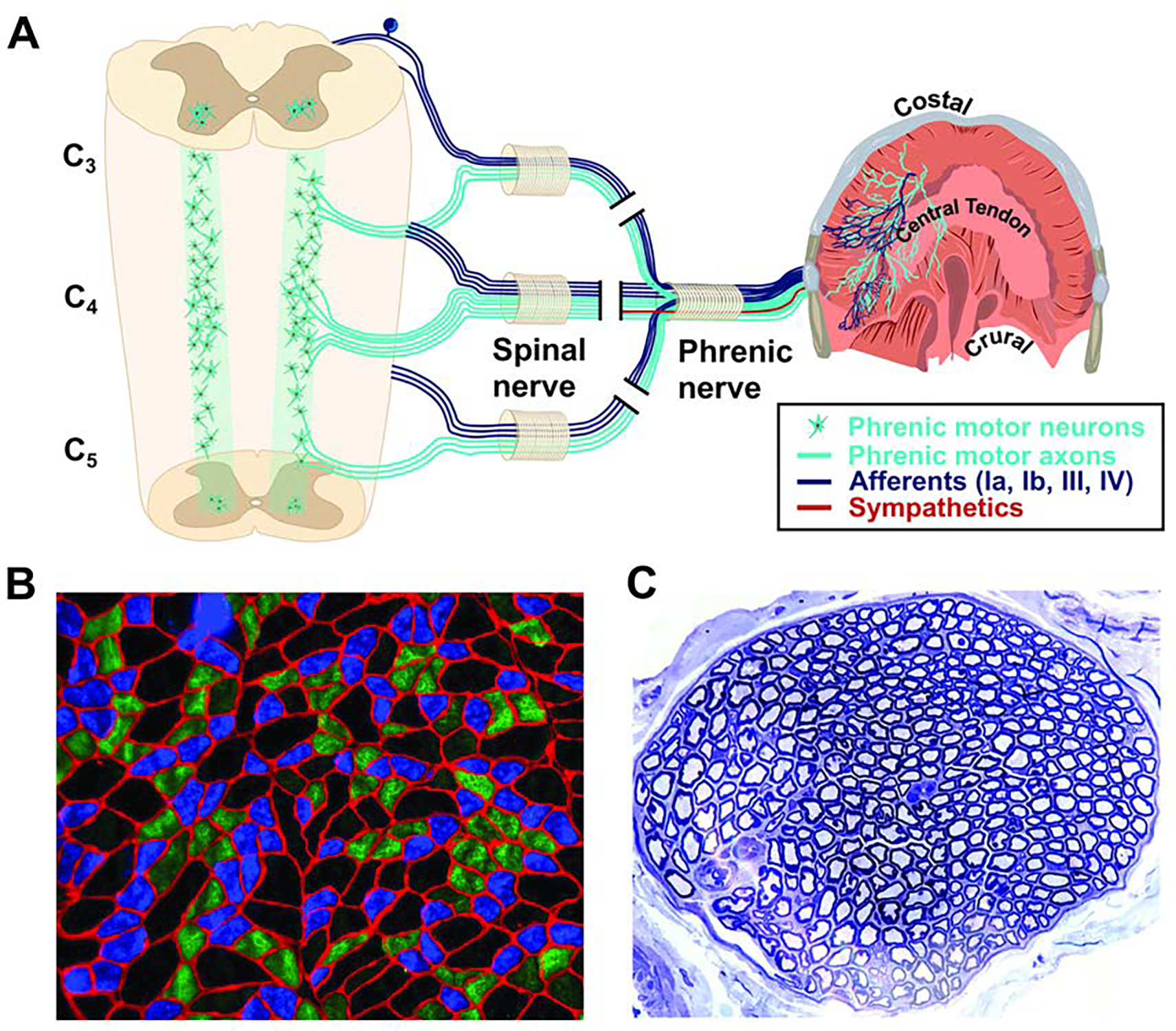
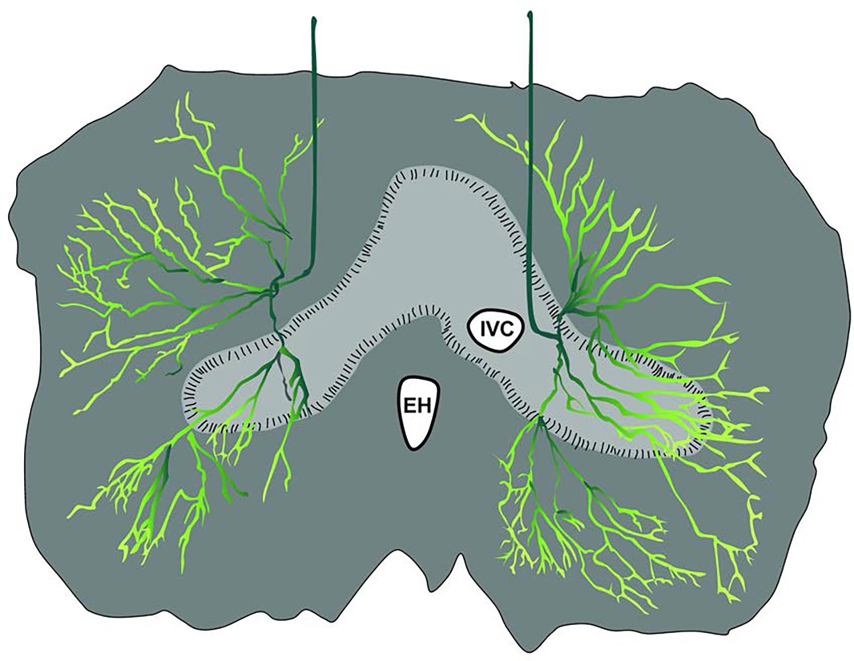
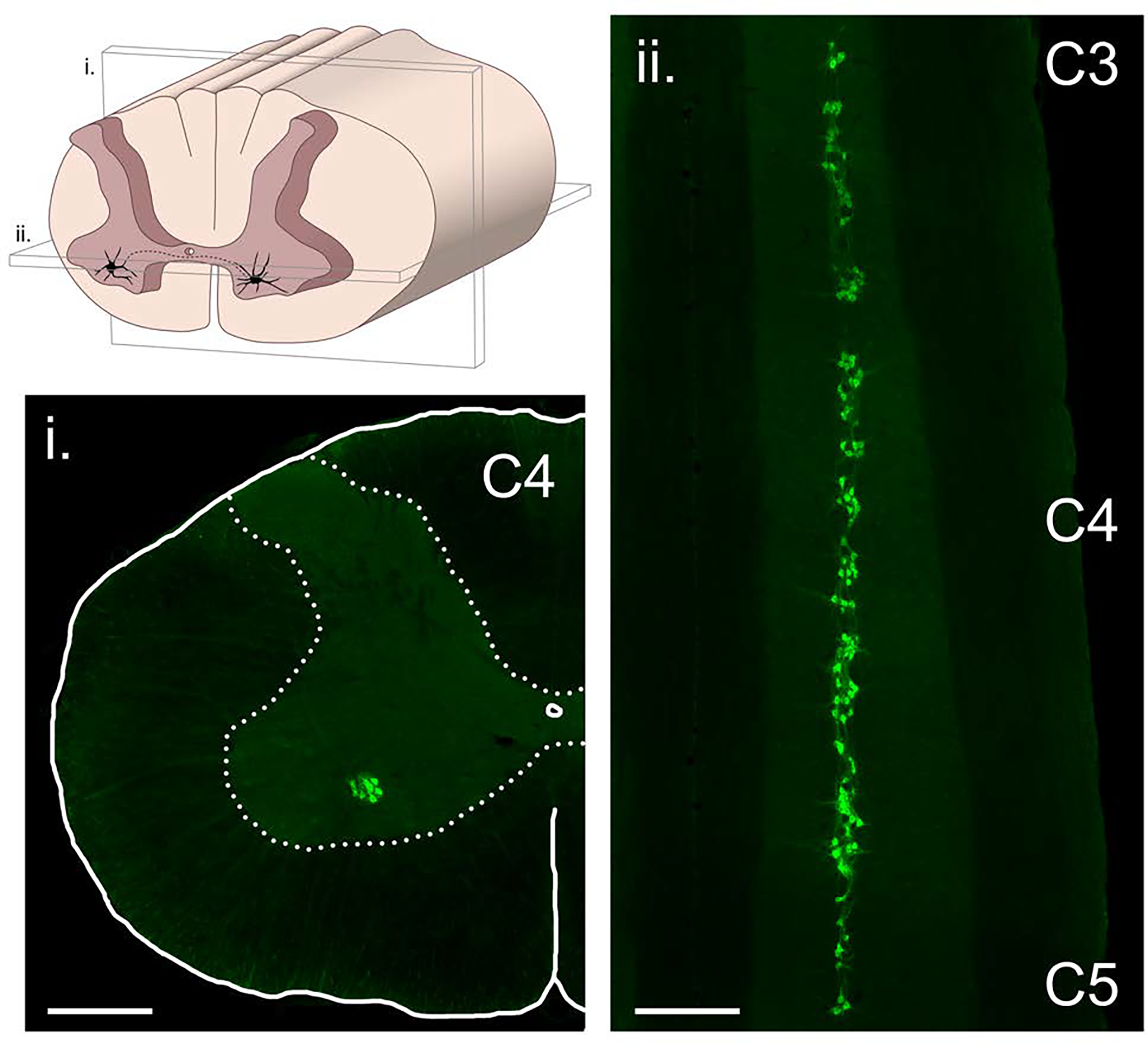
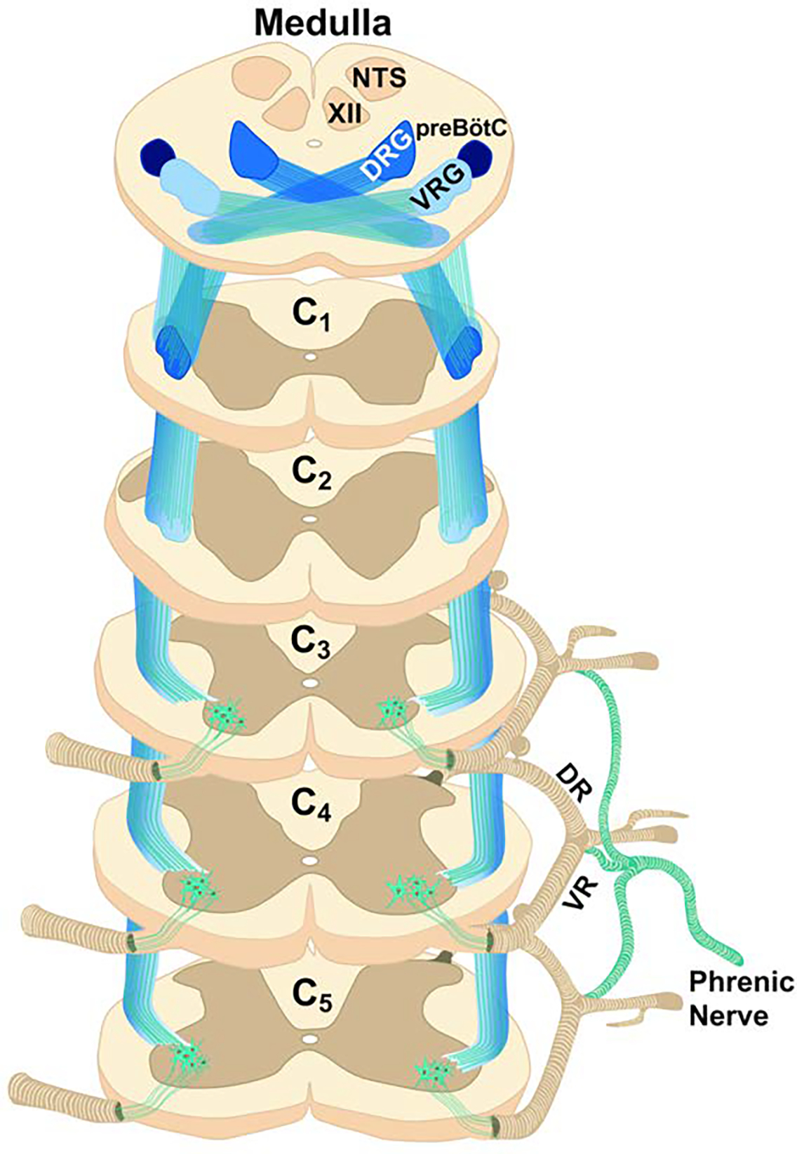
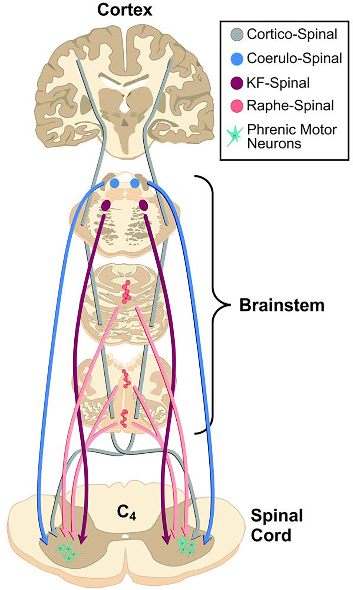
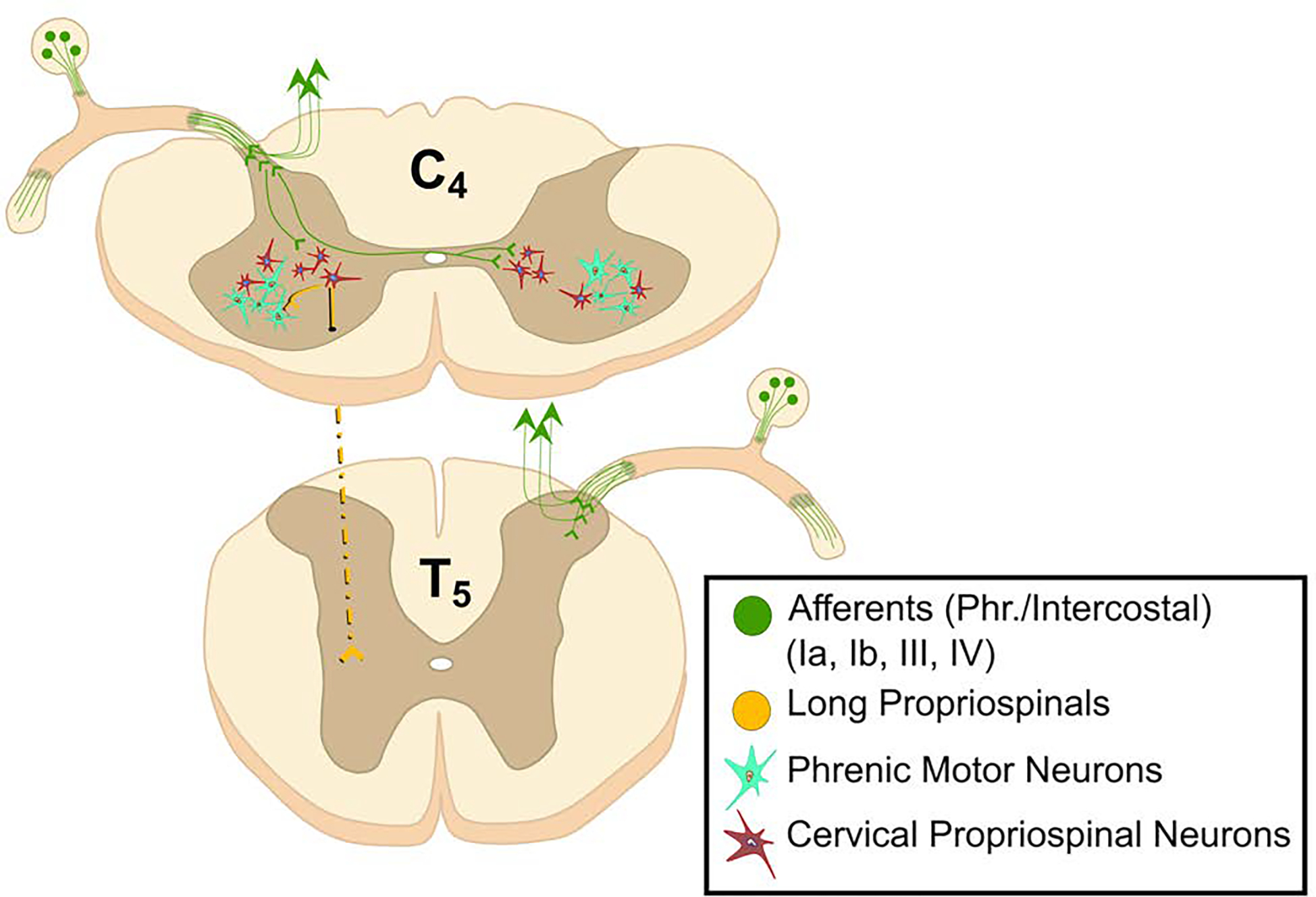
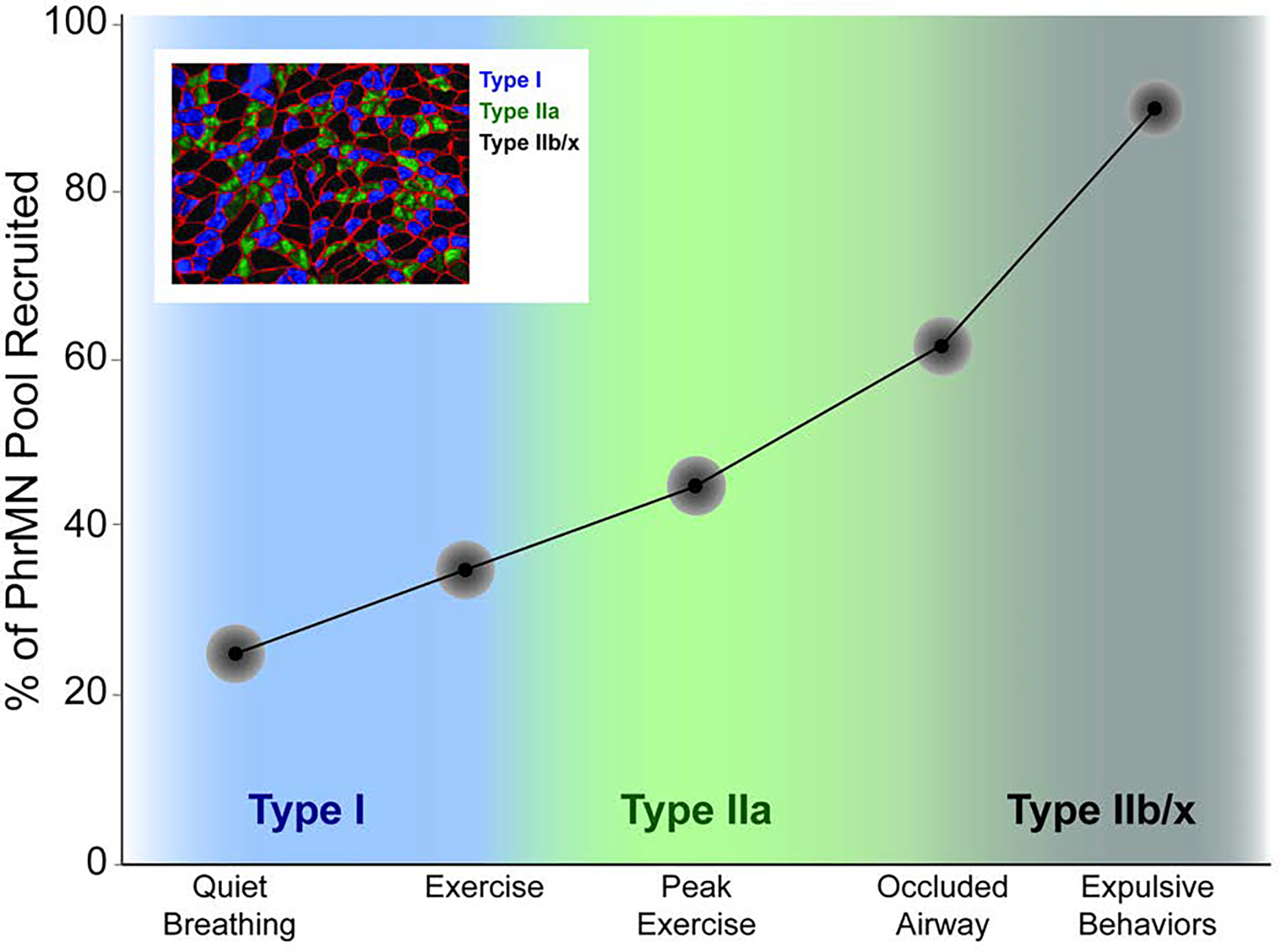
The phrenic neuromuscular system consists of the phrenic motor nucleus in the mid-cervical spinal cord, the phrenic nerve, and the diaphragm muscle. This motor system helps sustain breathing throughout life, while also contributing to posture, coughing, swallowing, and speaking. The phrenic nerve contains primarily efferent phrenic axons and afferent axons from diaphragm sensory receptors but is also a conduit for autonomic fibers. On a breath-by-breath basis, rhythmic (inspiratory) depolarization of phrenic motoneurons occurs due to excitatory bulbospinal synaptic pathways. Further, a complex propriospinal network innervates phrenic motoneurons and may serve to coordinate postural, locomotor, and respiratory movements. The phrenic neuromuscular system is impacted in a wide range of neuromuscular diseases and injuries. Contemporary research is focused on understanding how neuromuscular plasticity occurs in the phrenic neuromuscular system and using this information to optimize treatments and rehabilitation strategies to improve breathing and related behaviors.

Keywords: Breathing; Diaphragm; Interneuron; Motoneuron; Phrenic; Spinal.


Figures

Figure 1.
Figure 1.. The phrenic neuromuscular system.
A. The diaphragm is innervated via the phrenic nerve. The phrenic nerve contains phrenic motoneuron axons which innervate diaphragm myofibers, sensory afferent fibers from the diaphragm, and sympathetic post-ganglionic fibers. The phrenic motor nucleus extends from C3-C5 in humans. B. Example histology from the costal diaphragm illustrating mixed myofiber type (Type 1, slow oxidative, blue; Type IIa, intermediate fibers, green, Type IIb/x, fast glycolytic, black). Red indicates the myofiber membrane (laminin stain). C. Histological cross section of the phrenic nerve stained with toluidine blue. Large myelinated axons are clearly visible. The examples in B and C were obtained from an adult rat. Scale bars. B, 130 μm; C, 300 μm.
Figure 2.
Figure 2.. The distribution of phrenic nerve branches in the diaphragm.
The drawing depicts the thoracic surface, and is based on histological evaluation of the human diaphragm (An et al., 2012). The main phrenic nerve trunk diverges to form distinct anterior and posterior branches, with lateral branches of varying number. The primary branches subdivide to form smaller branches, thereby creating a “net” which innervates the entire diaphragm. EH: esophageal hiatus; IVC: inferior vena cava
Figure 3.
Figure 3.. The phrenic motoneuron pool.
The column of phrenic motoneurons extends from approximately C3 to C5. The drawing in the upper left panel illustrates the location of phrenic motoneurons in the anterior horn, and also the plane of section that the two histological images were taken from. The histological images were obtained using a retrograde neuronal tracer (cholera toxin, ß-subunit) applied intrapleurally to the diaphragm muscle in an adult rat model. Scale bars indicate 500 μm.
Figure 4.
Figure 4.. Direct, excitatory monosynaptic connection between the medulla and phrenic motoneurons.
The illustrated synaptic projections provide the primary “inspiratory drive” which depolarize phrenic motoneurons during inspiration, and thereby trigger diaphragm contraction. The axons of these projections travel in lateral and ventral cervical funiculi (the image illustrates the ventro-lateral funiculus). As noted in the text, PhrMNs also receive monosynaptic inhibitory inputs from the Bötzinger complex in the medulla. VRG: ventral respiratory group; DRG: dorsal respiratory group; XII: hypoglossal motor nucleus; NTS: nucleus of the solitary tract; preBôtC: pre-Bötzinger complex
Figure 5.
Figure 5.. Additional tracts innervating phrenic motor neurons.
The phrenic motor nucleus is innervated by corticospinal pathways as well as brainstem serotonergic and noradrenergic neurons.
Figure 6.
Figure 6.. The phrenic motor nucleus receives synaptic inputs from spinal cord interneurons.
These propriospinal neurons do not produce the fundamental respiratory rhythm of breathing but play a role in modulating the excitability of phrenic motoneurons and coordinating postural, locomotor and respiratory movements. These cells may also provide a neural substrate for promoting diaphragm motor recovery after spinal cord injury.
Figure 7.
Figure 7.. A model depicting recruitment of phrenic motoneurons during progressively more intense diaphragm contraction.
This model was developed by Sieck, Mantilla, and colleagues, and the image presented has been adapted from their work (Fogarty et al., 2018a). The model predicts that a relatively small amount of the total phrenic motoneuron pool (approximately 20%) is required for typical quiet breathing. This number increases during conditions such as intense exercise, but only approaches 100% during high force expulsive maneuvers.

References

    1. Alilain WJ & Goshgarian HG (2008). Glutamate receptor plasticity and activity-regulated cytoskeletal associated protein regulation in the phrenic motor nucleus may mediate spontaneous recovery of the hemidiaphragm following chronic cervical spinal cord injury. Exp Neurol 212: 348–357. - PMC - PubMed
    1. Allan DW & Greer JJ (1997). Embryogenesis of the phrenic nerve and diaphragm in the fetal rat. J Comp Neurol 382: 459–468. - PubMed
    1. Alvares TS, Revill AL, Huxtable AG, et al. (2014). P2Y1 receptor-mediated potentiation of inspiratory motor output in neonatal rat in vitro. J Physiol 592: 3089–3111. - PMC - PubMed
    1. An X, Yue B, Lee JH, et al. (2012). Intramuscular distribution of the phrenic nerve in human diaphragm as shown by Sihler staining. Muscle Nerve 45: 522–526. - PubMed
    1. Bains KNS, Kashyap S & Lappin SL (2021). Anatomy, Thorax, Diaphragm. StatPearls. Treasure Island (FL). - PubMed

LinkOut - more resources