. 2022 Aug 14;12(1):128.
doi: 10.1186/s13578-022-00867-7.

D-dopachrome tautomerase drives astroglial inflammation via NF-κB signaling following spinal cord injury

Affiliations

D-dopachrome tautomerase drives astroglial inflammation via NF-κB signaling following spinal cord injury

Hui Li et al. Cell Biosci. .

Abstract

Background: Reactive astrocytes are increasingly recognized as crucial regulators of innate immunity in degenerative or damaged central nervous system (CNS). Many proinflammatory mediators have been shown to drive inflammatory cascades of astrocytes through activation of NF-κB, thereby affecting the functional outcome of the insulted CNS. D-dopachrome tautomerase (D-DT), a newly described cytokine and a close homolog of proinflammatory macrophage migration inhibitory factor (MIF), has been revealed to share receptor and overlapping functional spectrum with MIF, but little is known about its roles in the neuropathological progression of the CNS and relevant regulatory mechanisms.

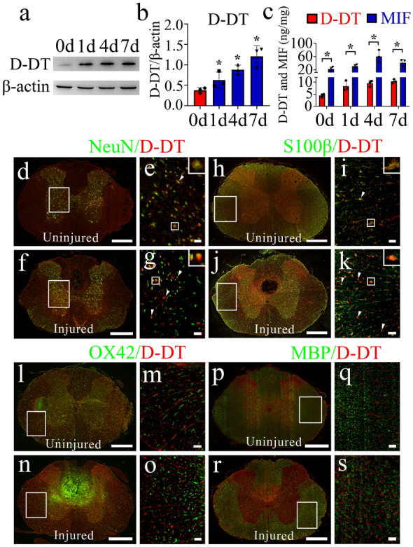
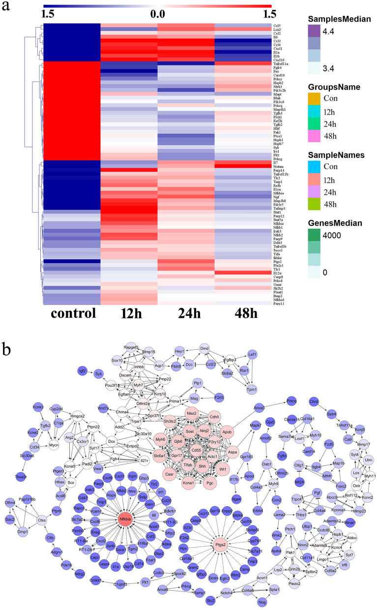
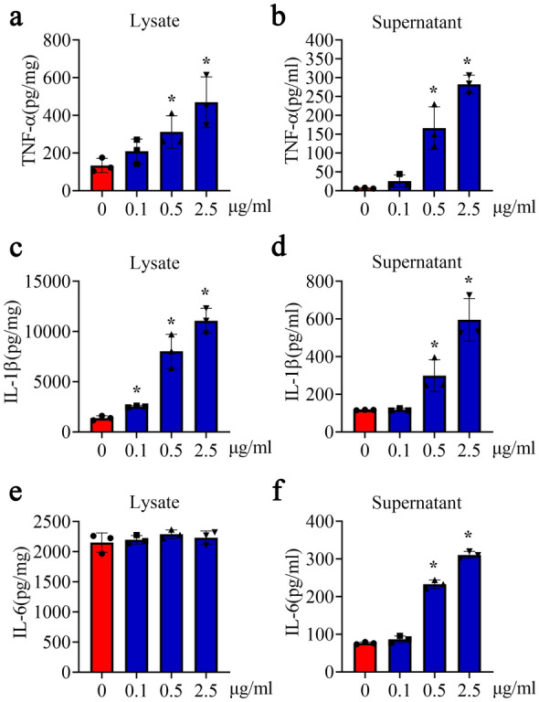
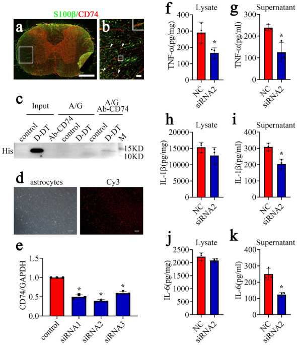
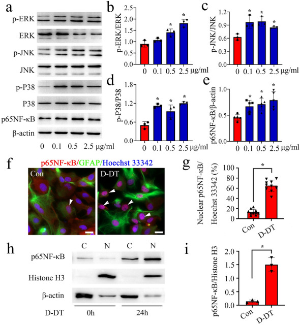
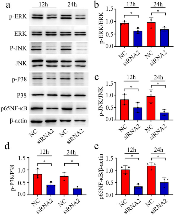
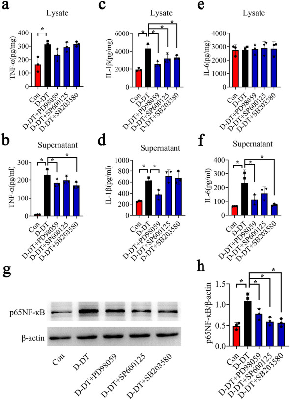
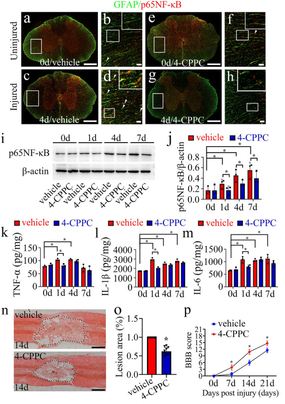
Results: D-DT protein levels were significantly elevated within neurons and astrocytes following SCI. Analysis of transcriptome profile revealed that D-DT was able to activate multiple signal pathways of astrocytes, which converged to NF-κB, a hub regulator governing proinflammatory response. Rat D-DT recombinant protein was efficient in inducing the production of inflammatory cytokines from astrocytes through interaction with CD74 receptor. Activation of mitogen-activated protein kinases (MAPKs) and NF-κB was observed to be essential for the transduction of D-DT signaling. Administration of D-DT specific inhibitor at lesion sites of the cord resulted in significant attenuation of NF-κB activation and reduction of the inflammatory cytokines following SCI, and accordingly improved the recovery of locomotor functions.

Conclusion: Collectively, D-DT is a novel proinflammatory mediator of astrocytes following SCI. Insights of its cell-specific expression and relevant proinflammatory mechanisms will provide clues for the control of CNS inflammation.

Keywords: Astrocyte; Central nervous system; D-DT; Inflammation; MAPKs; NF-κB; Spinal cord injury.


Conflict of interest statement

The authors have declared that no competing interests exist.

Figures

Fig. 1
Fig. 1
Examination of D-DT expression changes at lesion sites following rat SCI. a Western blot analysis of D-DT following cord contusion at 0d, 1d, 4d and 7d, respectively. b Quantification data as shown in a. Quantities were normalized to endogenous β-actin. c ELISA measurement of D-DT and MIF protein levels at lesion sites following SCI at 0d, 1d, 4d and 7d, respectively. d-s Immunostaining showed colocalization of D-DT with NeuN-positive neurons (d-g) and S100β-positive astrocytes (h-k), rather than with OX42-positive microglia (l-o) or MBP-positive oligodendrocytes (p-s) before or after SCI at 4d. Rectangle indicates region magnified. Arrowheads indicate the positive signals. Scale bars, 500 μm in (d), (f), (h), (j), (l), (n), (p) and (r); 50 μm in (e), (g), (i), (k), (m), (o), (q) and (s). Experiments were performed in triplicates. Error bars represent the standard deviation (*P < 0.05)
Fig. 2
Fig. 2
Expression profiling and gene network analysis of integrated DEGs in the astrocytes following stimulation with 1 µg/ml recombinant D-DT protein for 12, 24, and 48 h, respectively. a Heatmap and cluster dendrogram of integrated DEGs at 12, 24, and 48 h. The color scale shown at the top illustrates the relative expression level of the indicated mRNA across all samples: red denotes expression > 0 and blue denotes expression < 0. b A reconstructed gene network was created using the IPA on the basis of integrated DEGs
Fig. 3
Fig. 3
Determination of D-DT-induced inflammatory cytokines from astrocytes. The supernatants and lysates of astrocytes were determined by ELISA for the inflammatory cytokines TNF-α (a, b), IL-1β (c, d) and IL-6 (e, f) after cell stimulation with 0-2.5 µg/ml recombinant D-DT protein for 24 h. Experiments were performed in triplicates. Error bars represent the standard deviation (*P < 0.05)
Fig. 4
Fig. 4
D-DT promoted astrocytic production of inflammatory cytokines through interaction with CD74 receptor. a, b Immunofluorescence showed colocalization of CD74 with S100β-positive astrocytes in the spinal cord. Rectangle indicates region magnified. Arrowheads indicate the positive signals. c Binding assay of D-DT with CD74 receptor in the primary astrocytes. The immunoprecipitation was performed with anti-CD74 antibody (Ab-CD74), followed by detection of the components of the CD74-associated complexes with anti-His antibody. The control meant that the astrocytes were stimulated with 0.01 M PBS, and the D-DT indicated that the astrocytes were stimulated with 1 µg/ml recombinant D-DT protein with N-terminal His-tag. d Determination of siRNA transfection efficiency by Cy3 control. e Interference efficiency of three siRNA oligonucleotides for CD74 was measured by RT-PCR, and siRNA2 was used for the knockdown experiments. f-k Determination of TNF-α (f, g), IL-1β (h, i) and IL-6 (j, k) by ELISA from astrocytes following CD74 knockdown for 48 h and subsequent stimulation with 1.0 µg/ml recombinant D-DT protein for 24 h. Scrambles were used as control. Scale bars, 500 μm in (a), 50 μm in (b) and (d). Experiments were performed in triplicates. Error bars represent the standard deviation (*P < 0.05)
Fig. 5
Fig. 5
Effects of D-DT on the activation of intracellular MAPKs/NF-κB signaling in the astrocytes. a Western blot analysis of phosphorylation of ERK, P38, JNK kinase and p65NF-κB protein after stimulation of astrocytes with 0-2.5 µg/ml recombinant D-DT protein for 24 h. b–e Quantification data as shown in a. f Immunofluorescence showed the distribution of p65NF-κB in the cytoplasm and nucleus of astrocytes following treatment with 1 µg/ml recombinant D-DT for 24 h. Arrowheads indicate p65NF-κB-positive nucleus. g Quantification of p65NF-κB-positive nucleus co-stained with Hoechst 33,342 as shown in f. h Western blot analysis of p65NF-κB protein levels in the cytoplasm and nucleus following astrocytes treatment with 1 µg/ml recombinant D-DT for 24 h. i Quantification data as shown in h. Quantities were normalized to endogenous β-actin (cytoplasm) or Histone H3 (nucleus). Scale bar, 20 μm in f. Experiments were performed in triplicates. Error bars represent the standard deviation (*P < 0.05)
Fig. 6
Fig. 6
Effects of knockdown of CD74 receptor on the D-DT-mediated intracellular activation of MAPKs/NF-κB signaling in the astrocytes. a Western blot analysis of phosphorylation of ERK, P38, JNK kinase and p65NF-κB protein after siRNA2 knockdown of CD74 receptor for 48 h, prior to stimulation with 1 µg/ml recombinant D-DT protein for 12 and 24 h, respectively. b–e Quantification data as shown in a. Quantities were normalized to endogenous β-actin. Experiments were performed in triplicates. Error bars represent the standard deviation (*P < 0.05)
Fig. 7
Fig. 7
Inhibition of MAPKs attenuated D-DT-induced production of inflammatory cytokines from astrocytes. a–f ELISA determination of TNF-α (a, b), IL-1β (c, d) and IL-6 (e, f) in supernatant and lysate following astrocytes treatment with 1 µg/ml recombinant D-DT in the presence of 10 µM P38 (SB203580), 10 µM JNK (SP600125), or 10 µM ERK (PD98059) inhibitor for 24 h. g, h Western blot analysis of p65NF-κB protein levels following astrocytes treatment with 1 µg/ml recombinant D-DT in the presence of 10 µM P38 (SB203580), 10 µM JNK (SP600125), or 10 µM ERK (PD98059) inhibitor for 24 h. Experiments were performed in triplicates. Error bars represent the standard deviation (*P < 0.05)
Fig. 8
Fig. 8
D-DT inhibitor attenuates NF-κB signaling and ameliorates locomotor function following rat SCI. a–h Immunostaining showed colocalization of p65NF-κB with GFAP-positive astrocytes following injection of 8 µl of 100 mM vehicle or 4-CPPC at lesion sites of the contused cord at 0d and 4d. Rectangle indicates region magnified. Arrowheads indicate the positive signals. (i) Western blot analysis of p65NF-κB at lesion sites following treatment with vehicle or 4-CPPC inhibitor at 0d, 1d, 4d and 7d. j Quantification data as shown in (i). Quantities were normalized to endogenous β-actin. k–m ELISA assay of TNF-α (k), IL-1β (l) and IL-6 (m) following cord treatment with vehicle or 4-CPPC inhibitor at 0d, 1d, 4d, and 7d, respectively. n HE staining of the injured spinal cord at 14 d after injection of 8 µl of 100 mM vehicle or 4-CPPC inhibitor at lesion sites. o Quantification data as shown in n. p BBB score of hindlimb at 0d, 7d, 14d and 21d following intrathecal injection of 8 µl of 100 mM 4-CPPC or vehicle at the lesion sites, n = 6. Scale bars, 500 μm in (a), (c), (e), (g) and (n); 50 μm in (b), (d), (f) and (h). Experiments were performed in triplicates. Error bars represent the standard deviation (*P < 0.05)

Similar articles

Cited by

References

    1. Han RT, Kim RD, Molofsky AV, Liddelow SA. Astrocyte-immune cell interactions in physiology and pathology. Immunity. 2021;54(2):211–24. doi: 10.1016/j.immuni.2021.01.013. - DOI - PubMed
    1. Pekny M, Pekna M. Astrocyte reactivity and reactive astrogliosis: costs and benefits. Physiol Rev. 2014;94(4):1077–98. doi: 10.1152/physrev.00041.2013. - DOI - PubMed
    1. Colombo E, Farina C, Astrocytes Key regulators of neuroinflammation. Trends Immunol. 2016;37(9):608–20. doi: 10.1016/j.it.2016.06.006. - DOI - PubMed
    1. Yi W, Schluter D, Wang X. Astrocytes in multiple sclerosis and experimental autoimmune encephalomyelitis: Star-shaped cells illuminating the darkness of CNS autoimmunity. Brain Behav Immun. 2019;80:10–24. doi: 10.1016/j.bbi.2019.05.029. - DOI - PubMed
    1. Sofroniew MV. Astrocyte Reactivity: Subtypes, States, and Functions in CNS Innate Immunity. Trends Immunol. 2020;41(9):758–70. doi: 10.1016/j.it.2020.07.004. - DOI - PMC - PubMed

LinkOut - more resources