

Functions and mechanisms of protein disulfide isomerase family in cancer emergence
- PMID: 35965326
- PMCID: PMC9375924
- DOI: 10.1186/s13578-022-00868-6
Functions and mechanisms of protein disulfide isomerase family in cancer emergence
Abstract
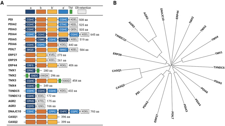
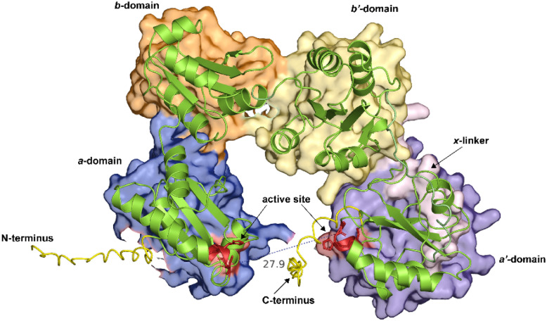
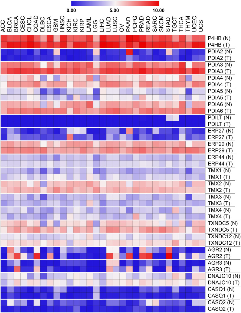
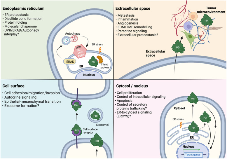
The endoplasmic reticulum (ER) is a multi-layered organelle that is essential for the synthesis, folding, and structural maturation of almost one-third of the cellular proteome. It houses several resident proteins for these functions including the 21 members of the protein disulfide isomerase (PDI) family. The signature of proteins belonging to this family is the presence of the thioredoxin domain which mediates the formation, and rearrangement of disulfide bonds of substrate proteins in the ER. This process is crucial not only for the proper folding of ER substrates but also for maintaining a balanced ER proteostasis. The inclusion of new PDI members with a wide variety of structural determinants, size and enzymatic activity has brought additional epitomes of how PDI functions. Notably, some of them do not carry the thioredoxin domain and others have roles outside the ER. This also reflects that PDIs may have specialized functions and their functions are not limited within the ER. Large-scale expression datasets of human clinical samples have identified that the expression of PDI members is elevated in pathophysiological states like cancer. Subsequent functional interrogations using structural, molecular, cellular, and animal models suggest that some PDI members support the survival, progression, and metastasis of several cancer types. Herein, we review recent research advances on PDIs, vis-à-vis their expression, functions, and molecular mechanisms in supporting cancer growth with special emphasis on the anterior gradient (AGR) subfamily. Last, we posit the relevance and therapeutic strategies in targeting the PDIs in cancer.
© 2022. The Author(s).
Conflict of interest statement
The authors declare that they have no competing interests.
Figures
Similar articles
-
Modulation of Protein Disulfide Isomerase Functions by Localization: The Example of the Anterior Gradient Family.Antioxid Redox Signal. 2024 Oct;41(10-12):675-692. doi: 10.1089/ars.2024.0561. Epub 2024 Apr 3. Antioxid Redox Signal. 2024. PMID: 38411504 Review.
-
Structures and functions of protein disulfide isomerase family members involved in proteostasis in the endoplasmic reticulum.Free Radic Biol Med. 2015 Jun;83:314-22. doi: 10.1016/j.freeradbiomed.2015.02.010. Epub 2015 Feb 17. Free Radic Biol Med. 2015. PMID: 25697777 Review.
-
The Protein Disulfide Isomerase Family: from proteostasis to pathogenesis.Biochim Biophys Acta Gen Subj. 2020 Feb;1864(2):129338. doi: 10.1016/j.bbagen.2019.04.003. Epub 2019 Apr 12. Biochim Biophys Acta Gen Subj. 2020. PMID: 30986509 Review.
-
Emerging roles of protein disulfide isomerase in cancer.BMB Rep. 2017 Aug;50(8):401-410. doi: 10.5483/bmbrep.2017.50.8.107. BMB Rep. 2017. PMID: 28648146 Free PMC article. Review.
-
Protein disulfide isomerase family mediated redox regulation in cancer.Adv Cancer Res. 2023;160:83-106. doi: 10.1016/bs.acr.2023.06.001. Epub 2023 Jul 18. Adv Cancer Res. 2023. PMID: 37704292 Free PMC article. Review.
Cited by
-
Biological mechanisms and clinical significance of endoplasmic reticulum oxidoreductase 1 alpha (ERO1α) in human cancer.J Exp Clin Cancer Res. 2024 Mar 8;43(1):71. doi: 10.1186/s13046-024-02990-4. J Exp Clin Cancer Res. 2024. PMID: 38454454 Free PMC article. Review.
-
Protein disulphide isomerase A4 as a potential biomarker for coronavirus disease 2019: Correlation with cytokine profiles and disease progression.Virulence. 2025 Dec;16(1):2508815. doi: 10.1080/21505594.2025.2508815. Epub 2025 May 26. Virulence. 2025. PMID: 40391685 Free PMC article.
-
The role of molecular subtypes and immune infiltration characteristics based on disulfidptosis-associated genes in lung adenocarcinoma.Aging (Albany NY). 2023 Jun 13;15(11):5075-5095. doi: 10.18632/aging.204782. Epub 2023 Jun 13. Aging (Albany NY). 2023. PMID: 37315289 Free PMC article.
-
Agr2 in cancer and beyond: unraveling its role during protein synthesis, ER stress, and as a predictive biomarker.Mol Cell Biochem. 2025 Jun 5. doi: 10.1007/s11010-025-05318-8. Online ahead of print. Mol Cell Biochem. 2025. PMID: 40471515 Review.
-
Pan-Inhibition of Protein Disulfide Isomerase Caused Cell Death through Disrupting Cellular Proteostasis in Pancreatic Ductal Adenocarcinoma Cells.Int J Mol Sci. 2023 Nov 17;24(22):16467. doi: 10.3390/ijms242216467. Int J Mol Sci. 2023. PMID: 38003657 Free PMC article.
References
Publication types
Grants and funding
LinkOut - more resources
Full Text Sources
