. 2022 Aug 14;23(1):39.
doi: 10.1186/s12865-022-00515-2.

NLRP3 inflammasome up-regulates major histocompatibility complex class I expression and promotes inflammatory infiltration in polymyositis

Affiliations

NLRP3 inflammasome up-regulates major histocompatibility complex class I expression and promotes inflammatory infiltration in polymyositis

Ping Xia et al. BMC Immunol. .

Abstract

Objective: This study was designed to investigate the role of the nucleotide-binding-domain -and leucine-rich repeat -containing (NLR) family, pyrin-domain-containing 3 (NLRP3) inflammasome in the pathogenesis of polymyositis (PM).

Methods: Immunochemistry was performed to analyze the NLRP3, caspase-1 and interleukin-1 beta (IL-1β) expression in the muscle tissue of PM patients. Rat model of PM and C2C12 cell were used to investigate the potential role of NLRP3 inflammasome in PM.

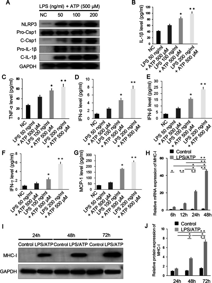
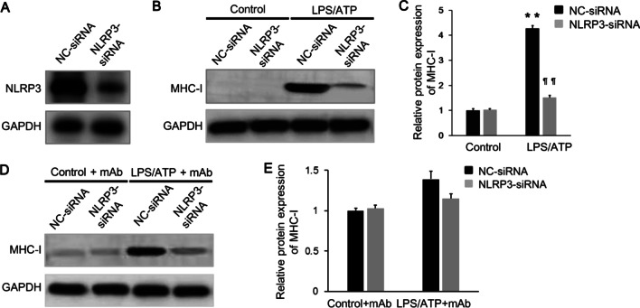
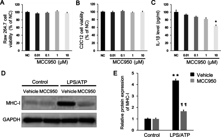
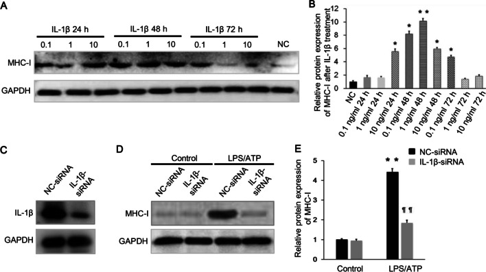
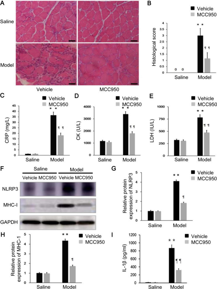
Results: The percentage of CD 68+ macrophages, and the expression levels of NLRP3, caspase-1 and IL-1β in the muscle tissue were elevated in 27 PM patients. LPS/ATP treatment resulted in activation of NLRP3 inflammasome and secretion of IL-1β as well as interferons (IFNs) and monocyte chemotactic protein-1 (MCP-1) in the Raw 264.7 macrophages. Meanwhile, LPS/ATP challenged activation of NLRP3 inflammasome induced overexpression of major histocompatibility complex class I (MHC-I), a key molecular of PM in the co-cultured C2C12 cells. The effect was decreased by treatment of NLRP3 inflammasome inhibitor MCC950 or siRNA of NLRP3 inflammasome. These findings suggested certain levels of IL-1β rather than IFNs up-regulated MHC-I expression in C2C12 cells. IL-1β blockade using neutralizing IL-1β monoclonal antibody or siRNA of IL-1β suppressed MHC-I overexpression. In vivo, NLRP3 inflammasome inhibition by MCC950 reduced the expression of NLRP3, IL-1β and MHC-I in the muscle tissue of PM modal rats. Also, it attenuated the intensity of muscle inflammation as well as the CRP, CK, and LDH levels in the serum.

Conclusion: NLRP3/caspase-1/IL-1β axis may play an important role in the development of PM. Inhibition of NLRP3 activation may hold promise in the treatment of PM.

Keywords: Autoimmune diseases; Inflammation; MCC950; Major histocompatibility complex class I; NLRP3 inflammasome; Polymyositis.


Conflict of interest statement

The authors declare that the research was conducted in the absence of any commercial or financial relationships that could be constructed as a potential conflict of interest.

Figures

Fig. 1
Fig. 1
The level of inflammation is increased in the PM patients and the NLRP3/caspase-1/ IL-1β axis is active in their muscle samples. A Immunohistochemistry analysis showed higher percentage of CD68+ cells (solid black arrows) in the muscle biopsies in the PM patients than in the controls. B Immunofluorescence staining showed positive MHC-I staining (grade+ ~ ++) in the PM patients, and negative in the controls (grade−). C Immunohistochemistry analysis of the muscle biopsies showed higher expression of NLRP3, caspase-1 and IL-1β (solid black arrows) in the PM patients than in the controls. **p < 0.001; scale bars, 50 μm (original magnification, ×400)
Fig. 2
Fig. 2
LPS/ATP stimulation activates NLRP3/caspase-1/IL-1β axis and promotes MHC-I expression. A Western blot analysis showed increased expression of NLRP3, pro-caspase-1, cleaved caspase-1, pro-IL-1β and mature IL-1β in the Raw 264.7 macrophages under LPS/ATP stimulation. B, C ELISA analysis showed that the expression of IL-1β and TNF-α in the Raw 264.7 macrophages were up-regulated by LPS/ATP. DG ELISA analysis showed elevated levels of IFNs and MCP-1 produced by the stimulated macrophages. H The mRNA expression of MHC-I in the co-cultured C2C12 cells was increased under LPS/ATP stimulation and peaked at 48 h. I, J The protein expression of MHC-I in the co-cultured C2C12 cells was increased under LPS/ATP stimulation and peaked at 72 h. *p < 0.01, **p < 0.001, compared with control group or compared between the groups connecting with the line
Fig. 3
Fig. 3
Genetic knockdown of NLRP3 suppresses MHC-I overexpression in C2C12 cells. A Raw 264.7 macrophages were transfected with siRNA targeting NLRP3, and the transfection efficiency was measured by western blot analysis. B, C Raw 264.7 macrophages pretreated with NLRP3-siRNA were stimulated with 200 ng/ml LPS + 500 μM ATP, and then the expression of MHC-I in the co-cultured C2C12 cells was significantly reduced compared with those pretreated with NC-siRNA, as revealed by western blot. D, E The stimulated Raw 264.7 macrophages were pretreated with NLRP3-siRNA or NC-siRNA, and the co-cultured C2C12 cells were added with neutralizing IL-1β monoclonal antibody. No differences in the expression of MHC-I in the co-cultured C2C12 cells were found between those groups stimulated with 200 ng/ml LPS + 500 μM ATP or with PBS, as showed by western blot. **p < 0.001, compared with control group; ¶¶p < 0.001, compared with before treatment
Fig. 4
Fig. 4
Pharmacological inhibition of NLRP3 inflammasome using MCC950 effectively suppresses MHC-I overexpression in C2C12 cells. A, B Cell viability assay revealed no significant cytotoxicity of MCC950 to the Raw 264.7 cells and C2C12 cells, at the dosage from 0.01 to 10 μM. C LPS/ATP stimulated Raw 264.7 macrophages were treated by MCC950 with dosage from 0.01 to 10 μM, and the IL-1β level in the cell supernatant was significantly decreased at the dosage of 10 μM. D, E LPS/ATP stimulated Raw 264.7 macrophages were pretreated by 10 μM MCC950, and the MHC-I expression in the co-cultured C2C12 cells was significantly suppressed as showed by western blot. *p < 0.01, **p < 0.001, compared with control group; ¶¶p < 0.001, compared with before treatment
Fig. 5
Fig. 5
IL-1β is crucial for MHC-I up-regulation in C2C12 cells. A, B Recombinant IL-1β was added into the C2C12 cells. The expression of MHC-I in C2C12 cells peaked when treated by 1 ng/ml IL-1β for 48 h (n = 5). C Raw 264.7 macrophages were transfected with siRNA targeting IL-1β, and the transfection efficiency was measured by western blot analysis. D, E LPS/ATP stimulated Raw 264.7 macrophages were pretreated with IL-1β-siRNA or NC-siRNA, and the MHC-I expression in the co-cultured C2C12 cells was significantly lower with the former than with the latter pretreatment, as analyzed by western blot. *p < 0.01, **p < 0.001, compared with control group; ¶¶p < 0.001, compared with before treatment
Fig. 6
Fig. 6
MCC950 alleviates the intensity of inflammation and MHC-I expression in PM model rats. A, B The muscle section samples of PM modal rats showed a substantial reduction in muscle histological inflammation scores after treated by MCC950. CE Serum CRP, CK, and LDH levels were significantly reduced after MCC950 treatment in PM modal rats. FI The expression of NLRP3, MHC-I, and IL-1β were effectively reduced in the muscle tissue of PM model rats by MCC950 treatment. **p < 0.001, compared with control group; ¶p < 0.01, ¶¶p < 0.001, compared with before treatment. Scale bars, 50 μm (original magnification, ×400)

Similar articles

Cited by

References

    1. Selva-O’Callaqhan A, Pinal-Fernandez I, Trallero-Araquás E, Milisenda JC, Grau-Junyent JM, Mammen AL. Classification and management of adult inflammatory myopathies. Lancet Neurol. 2018;17:816–828. doi: 10.1016/S1474-4422(18)30254-0. - DOI - PMC - PubMed
    1. Mammen AL. Dermatomyositis and polymyositis: Clinical presentation, autoantibodies, and pathogenesis. Ann N Y Acad Sci. 2010;1184:134–153. doi: 10.1111/j.1749-6632.2009.05119.x. - DOI - PubMed
    1. Helmers SB, Bruton M, Loell I, Ulfgren AK, Gracie AJ, McInnes IB, et al. Expression of interleukin-18 in muscle tissue of patients with polymyositis or dermatomyositis and effects of conventional immunosuppressive treatment. Rheumatology. 2018;57:2149–2157. doi: 10.1093/rheumatology/key222. - DOI - PubMed
    1. Milone M. Diagnosis and management of immune-mediated myopathies. Mayo Clin Proc. 2017;92:826–837. doi: 10.1016/j.mayocp.2016.12.025. - DOI - PubMed
    1. Zhang SX, Wang J, Sun HH, Zhang JQ, Liu GY, Luo J, He P-F, Li X-F. Circulating regulatory T cells were absolutely decreased in dermatomyositis/polymyositis patients and restored by low-dose IL-2. Ann Rheum Dis. 2019;0:1–3. - PubMed