. 2023 Feb 1;31(2):471-486.
doi: 10.1016/j.ymthe.2022.08.005. Epub 2022 Aug 13.

DDX5 mRNA-targeting antisense oligonucleotide as a new promising therapeutic in combating castration-resistant prostate cancer

Affiliations

DDX5 mRNA-targeting antisense oligonucleotide as a new promising therapeutic in combating castration-resistant prostate cancer

Thi Khanh Le et al. Mol Ther. .

Abstract

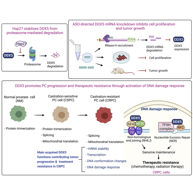
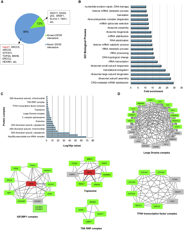
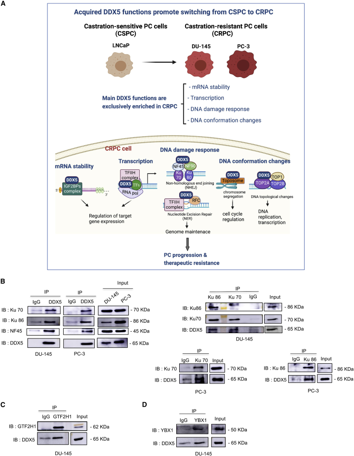
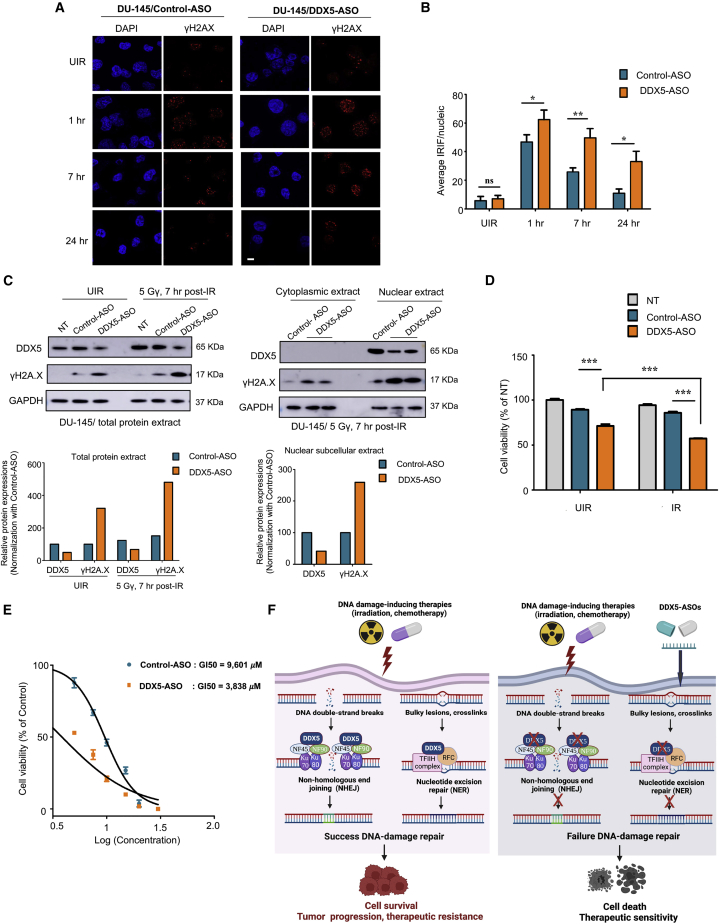
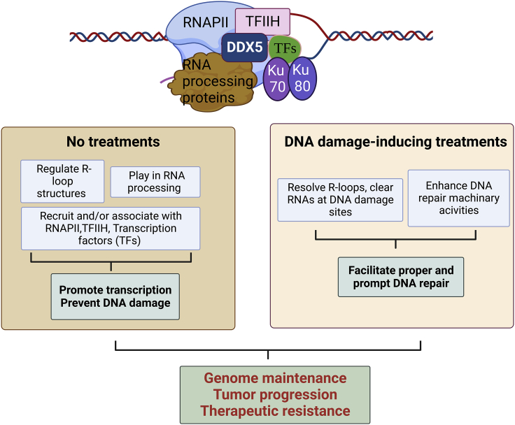
The heat shock protein 27 (Hsp27) has emerged as a principal factor of the castration-resistant prostate cancer (CRPC) progression. Also, an antisense oligonucleotide (ASO) against Hsp27 (OGX-427 or apatorsen) has been assessed in different clinical trials. Here, we illustrate that Hsp27 highly regulates the expression of the human DEAD-box protein 5 (DDX5), and we define DDX5 as a novel therapeutic target for CRPC treatment. DDX5 overexpression is strongly correlated with aggressive tumor features, notably with CRPC. DDX5 downregulation using a specific ASO-based inhibitor that acts on DDX5 mRNAs inhibits cell proliferation in preclinical models, and it particularly restores the treatment sensitivity of CRPC. Interestingly, through the identification and analysis of DDX5 protein interaction networks, we have identified some specific functions of DDX5 in CRPC that could contribute actively to tumor progression and therapeutic resistance. We first present the interactions of DDX5 and the Ku70/80 heterodimer and the transcription factor IIH, thereby uncovering DDX5 roles in different DNA repair pathways. Collectively, our study highlights critical functions of DDX5 contributing to CRPC progression and provides preclinical proof of concept that a combination of ASO-directed DDX5 inhibition with a DNA damage-inducing therapy can serve as a highly potential novel strategy to treat CRPC.

Keywords: DDX5; DNA damage response (DDR); Hsp27; antisense oligonucleotides (ASOs); castration-resistant prostate cancer (CRPC); protein interactions; therapeutic ressistance.


Conflict of interest statement

Declaration of interests The authors declare to have no financial, personal, or professional competing interest and no conflict of interest.

Figures

None
Graphical abstract
Figure 1
Figure 1
Hsp27 positively regulates DDX5 expression through preventing DDX5 protein from proteasomal degradation (A) Western blotting (WB) analyzing the protein expression levels of DDX5 and Hsp27 in LNCaP-mock versus LNCaP-Hsp27 (overexpressed Hsp27) shows that DDX5 expression is higher in LNCaP-Hsp27 compared with the LNCaP-mock. (B) WB analyzing the protein expressions of DDX5 in the LNCaP and PC-3 cells transfected with OGX-427 (an Hsp27-targeting ASO) or control-ASO reveals that DDX5 protein expression decreased dramatically upon Hsp27 depletion. (C) Hsp27 does not regulate DDX5 at transcriptional level. qRT-PCR analysis showed that DDX5 mRNA levels are similar in both LNCaP-mock and LNCaP-Hsp27 cells. The mRNA levels of both DDX5 and Hsp27 are evaluated by qRT-PCR analysis using the PC-3 cells transfected with OGX-427 or control-ASO. The DDX5 mRNA level is constant upon Hsp27 knockdown. Sample t test was performed to compare the relative mRNA levels between two samples: LNCaP-mock versus LNCaP-Hsp27, OGX-427 versus control-ASO-transfected PC-3 cells. ∗∗p < 0.01; ns, non-significant. (D) Confirmation of the interaction between Hsp27 and DDX5. DDX5 IP followed by WB analysis showed that Hsp27 was present in the IP using anti-DDX5 antibody. (E and F) DDX5 stability is controlled by the proteasome pathway in PC. Proteasome inhibition using either 10 μM MG132 or 100 nM bortezomib induces accumulation of DDX5 protein at the indicated times (E). Proteasome inhibitor combined with an inhibitor of de novo protein synthesis, CHX (cycloheximide), conserves DDX5 protein levels (F). (G) Hsp27 prevents DDX5 from proteasome degradation. PC-3 cells were transfected with OGX-427 or control-ASO at 150 nM concentration for 2 days, followed by incubation with or without 100 nM bortezomib for 24 h. WB analysis shows that the proteasome inhibitor bortezomib can reverse the downregulation of DDX5 induced by OGX-427. (H) DDX5 does not control Hsp27 protein abundance. WB analyses of the DDX5 and Hsp27 expression upon either Hsp27 knockdown by OGX-427 or DDX5 downregulation by DDX5-ASO in PC-3 cells. Hsp27 protein expression is stable upon DDX5 depletion.
Figure 2
Figure 2
DDX5 overexpression is associated with tumor progression and CRPC as clinically relevant (A) DDX5 expression is significantly higher in prostate cancer (PC) compared with benign prostatic hyperplasia (BPH). (B) representative images of DDX5 staining show increased DDX5 expression in PC specimens compared with BPH. (C) results show a tendency for DDX5 expression to increase with increasing Gleason grade. (D) Representative images of DDX5 staining show increased staining intensity in Gleason grade 5 specimens. (E) The graph and the representative IHC images show that DDX5 expression is extremely higher in castration-resistant prostate cancer (CRPC) patients than in the hormone naive and neoadjuvant hormone therapy (NHT) tumors. (F) Representative images of DDX5 staining show a remarkable increase of DDX5 staining expression in CRPC specimens. (G) DDX5 is overexpressed in CR cells (DU-145 and PC-3) compared with CS cells (LNCaP). The DDX5 protein level was analyzed by western blot using total cell lysate from three different prostate cell lines (LNCaP, DU-145, and PC-3). DDX5 protein level was normalized with GAPDH, which was also used as loading control. Quantification illustrates that DDX5 expression levels in both CR lines are at about three times as high as in CS cells. (H) Kaplan-Meier RFS curves in the TCGA PRAD public samples. High expression level of DDX5 mRNAs is positively correlated with poor recurrence-free survival (RFS). According to the DDX5 mRNA levels, samples were classified into two groups, DDX5 high and DDX5 low, which have 10-year RFS rates of 34% and 63%, respectively in a pool of 490 informed patients. ∗p < 0.05; ∗∗p < 0.01; ∗∗∗p < 0.001; ∗∗∗∗p < 0.0001; ns, non-significant.
Figure 3
Figure 3
ASO-directed DDX5 downregulation inhibits cell proliferation and PC-3 xenograft-tumor growth (A–C) ASO screenings aim to define ASOs that inhibit DDX5 protein expression. Both hASO51 (human ASO) and hmASO3 (human/mouse) decrease DDX5 expression in a dose-dependent manner. (D) DDX5 knockdown by both hASO51 and hmASO3 significantly inhibits cell proliferation. The 3-(4,5-dimethylthiazol-2-yl)-2,5-diphenyl-2H-tetrazolium bromide (MTT) assay was done with biological triplication, and the data were analyzed using an independent sample t test. ∗∗∗p < 0.001. (E) Mean (± SE) growth curve of PC-3 xenografted tumors (∗∗∗p < 0.001, using repeated measures ANOVA). Male BALB/c nude mice harboring PC-3-derived xenografted tumor (7 mice/group) were daily intraperitoneally injected with 12.5 mg/kg control-ASO or DDX5-ASO (hmASO3) for 5 weeks. Tumor volume (mm3) was measured weekly using a caliper in three perpendicular dimensions (X = width, Y = length, Z = depth) and calculated following the formula X ×Y × Z × 0.5236. The group treated with DDX5-ASO had a delay in tumor growth compared with the group of control-ASO. (F) Population pharmacodynamic modeling demonstrates that DDX5-ASO treatment significantly inhibits the tumor growth compared with the control group. The left and middle panels present the goodness of fit and predictive check (VPC) plot. Overall, these two plots indicate that the base model provided an acceptable fit to the observed data. Left panel: Individual predicted volumes versus observed volumes. Points represent individual predicted volumes plotted against observed volumes and are distributed evenly around the identity line, indicating that no major bias was observed across predicted values. Middle panel: Results of the scatter visual predictive check from 500 model-generated datasets. Solid triangles represent the observed volumes and are almost within the 95% prediction interval of the model, demonstrating that the model is able to reproduce the pharmacodynamic (PD) observations adequately. Right panel: Model prediction of tumor growth kinetics, depending on the received treatment, using the final model. Individuals for the mice treated with DDX5-ASO showed a 20% decrease in maximum tumor volume. SCR: scrambled oligonucleotide; NT: non- treated; TSmax, individual carrying capacity (predicted maximum tumor volume).
Figure 4
Figure 4
Functional bioinformatics analyzing DDX5 interaction proteins identified in the study (A) The pie chart shows the proportion of the novel DDX5-interacting proteins (87.9%) and the identified interactions characterized as known DDX5-interacting proteins in the string database (12.1%). (B) Gene ontology biological process (GO-BP) enrichment analyses of the DDX5 interactome were performed using BiNGO (hypergeometric test, Benjamini and Hochberg false discovery rate [FDR] correction, p = 0.005, biological process). The bar chart shows the top functions ranked by the fold enrichment score. (C) CORUM enrichment analysis allows identifying experimentally proven human protein complexes associated with DDX5. (D) Network modeling of the top enriched protein complexes and DDX5. Green nodes represent proteins found in the DDX5 interactome obtained in the study, while gray nodes mean proteins were not identified. Bordered edges represent interactions with DDX5 annotated in published PPI databases. All of the protein members of the IGF2BP1 complex were found in the DDX5 interactions. A solid connection between DDX5 and the toposome was observed since most of the protein components of the toposome associate with DDX5 in the study (SSRP1 did not appear in the DDX5 interactions, but it was annotated to interact with DDX5 in string db). We showed the rigid association of DDX5 with the general transcription factor complex GTFIIH (or TFIIH), which is composed of the core complex (GTF2H1, GTF2H2, GTF2H3, GTF2H4, ERCC2, ERCC3) and the CAK (CDK7, CCNH, MNAT1). DDX5 could modulate transcription through the 7SK RPN complex because all of its constructive proteins (CDK9, HEXIM1, CCNT1, and LARP7) interact with DDX5.
Figure 5
Figure 5
Acquired DDX5 functions lead to switching from castration sensitivity to castration resistance of PC (A) The scheme describes the hypothesis about how DDX5 can promote PC progression and therapeutic resistance based on functional analyses of DDX5 interaction network. DDX5 could drive PC progression and therapeutic resistance through playing roles in DNA damage response, mRNA stabilization, transcription, and DNA conformation changes since these cellular processes are exclusively enriched in the DDX5 PPI obtained in CRCP. In particular, DDX5 regulates mRNA stability via interacting with IGF2BP complex, thereby modulating expression of target genes. DDX5 also regulates gene expression by monitoring transcription initiation via associating with the basal transcription complex TFIIH and transcription factors (TFs). DDX5 functions in non-homologous end joining (NHEJ) via interacting with the Ku70/Ku80 and NF45/NF90 complexes. DDX5 also interacts with the TFIIH complex and RFC proteins (RFC1, RFC3, RFC5) to participate in nucleotide excision repair (NER). DDX5 may regulate the cell cycle through its interaction with the toposome complex acting in chromosome segregation. Through binding to different topoisomerase enzymes such as TOP2A, TOP2B, and TOP1, DDX5 can modulate the DNA topological changes, thereby monitoring DNA replication and transcription. (B) IP using anti-DDX5 Ab followed by WB with anti-Ku70 Ab, anti-Ku80 Ab, and anti-NF45 Ab shows the presence of Ku70, Ku80, and NF45 in the DDX5 complexes in both CR DU-145 and PC-3 cells (left above panel). The reverse IP using anti-Ku70 Ab and anti- Ku80 Ab followed by WB using anti-DDX5 Ab confirmed the interaction of DDX5 with the Ku70/Ku80 heterodimer (right above and below panel). (C and D) Similarly, the IP using anti-GTF2H1 Ab and anti-YBX1 Ab combined with WB using anti-DDX5 Ab confirmed the association of DDX5 with GTF2H1 (C) and YBX1 (D).
Figure 6
Figure 6
DDX5 promotes therapeutic resistance of CRPC through activation of DNA repair (A and B) DDX5 downregulation reduces DNA damage repair efficiency by immunofluorescent (IF) and irradiation induced foci (IRIF) counting. The DU-145 cells were transfected with 150 nM DDX5-ASO (hmASO3) or control-ASO, and 3 days later, the cells were exposed to 5 Gy IR and further cultured for various time periods (UIR = unirradiated, 1, 7, and 24 h). IF staining using anti-γH2AX antibody was carried out (A). The graph shows a higher number of the IRIF in DDX5-ASO-treated cells compared with the control-ASO-transfected cells (B). (C) WB analysis shows that DDX5 depletion defects the DNA repair process. DU-145 cells transfected with the DDX5-ASO (hmASO3) or control-ASO (150 nM) were exposed to 5 Gy IR and further cultured for 7 h. The total protein extract or compartment extracts including cytoplasmic and nuclear extracts were subjected to WB analysis using anti-DDX5 Ab, anti-γH2AX Ab, and GAPDH used as a loading control. (D and E) DDX5 knockdown enhances cell sensitivity to DNA damage stress stimuli such as irradiation (D) and cisplatin (E). The DU-145 cells were transfected with DDX5-ASO for 2 days, treated with either irradiation or cisplatin, and subjected to cell proliferation evaluation using MTT assay after 2 days. The MTT tests were performed with triplication. DDX5-ASO can lower GI50 of cisplatin up to nearly three times. (F) In DDX5-overexpressed PC cells, DDX5 facilitates two main DNA repair pathways (NHEJ, NER), ensuring the proper DNA repair upon DNA damage-inducing therapies such as irradiation and chemotherapy, thereby promoting cell survival, PC development, and therapeutic resistance (left panel). Conversely, DDX5-downregulated PC cells failed to repair DNA lesions; therefore, they are sensitive to treatments (right panel). ∗p < 0.05; ∗∗p < 0.01; ∗∗∗p < 0.001; ns, non-significant.
Figure 7
Figure 7
The scheme illustrates a hypothesis picture of how DDX5 can promote tumor progression and therapeutic resistance via playing as a cross-talk among different signaling pathways such as transcription, RNA processing, and DNA repair in CRPC The increased R-loop formation frequently occurs in accessible chromatin regions of highly transcribed genes located in gene-rich areas that are more sensitive to DNA damage-inducing therapies than the other parts of the genome. Without any treatments, DDX5 mediates R-loops structure, recruits and/or binds with RNAPII, transcription initiation complex (TFIIH), and other transcription factors (TFs), and participates in coupling transcription-RNA process, thereby facilitating transcription process, preventing DNA damages, and promoting tumor development. On the other hand, upon DNA damage-inducing therapies, DDX5 can immediately facilitate DNA repair at the active transcription sites either via clearance of RNA transcripts of R-loop or enhancing the recruitment and/or activity of the DNA repair machinery (Ku complex, TFIIH complex), ensuring proper and rapid DNA repair, resulting in genomic maintenance and tolerance enhancement of cancer cells to therapies.

Similar articles

Cited by

References

    1. Bray F., Ferlay J., Soerjomataram I., Siegel R.L., Torre L.A., Jemal A. Global cancer statistics 2018: GLOBOCAN estimates of incidence and mortality worldwide for 36 cancers in 185 countries. CA. Cancer J. Clin. 2018;68:394–424. - PubMed
    1. Katsogiannou M., Ziouziou H., Karaki S., Andrieu C., Henry de Villeneuve M., Rocchi P. The hallmarks of castration-resistant prostate cancers. Cancer Treat. Rev. 2015;41:588–597. - PubMed
    1. Rocchi P., Jugpal P., So A., Sinneman S., Ettinger S., Fazli L., Nelson C., Gleave M. Small interference RNA targeting heat-shock protein 27 inhibits the growth of prostatic cell lines and induces apoptosis via caspase-3 activation in vitro. BJU Int. 2006;98:1082–1089. - PubMed
    1. Rocchi P., Beraldi E., Ettinger S., Fazli L., Vessella R.L., Nelson C., Gleave M. Increased Hsp27 after androgen ablation facilitates androgen-independent progression in prostate cancer via signal transducers and activators of transcription 3-mediated suppression of apoptosis. Cancer Res. 2005;65:11083–11093. - PubMed
    1. Rocchi P., So A., Kojima S., Signaevsky M., Beraldi E., Fazli L., Hurtado-Coll A., Yamanaka K., Gleave M. Heat shock protein 27 increases after androgen ablation and plays a cytoprotective role in hormone-refractory prostate cancer. Cancer Res. 2004;64:6595–6602. - PubMed

Publication types

MeSH terms