Case Reports
. 2022 Jul 28:12:797275.
doi: 10.3389/fonc.2022.797275. eCollection 2022.

Uterine perivascular epithelioid tumors (PEComas) with lung metastasis showed good responses to mTOR and VEGFR inhibitors: A case report

Affiliations
Case Reports

Uterine perivascular epithelioid tumors (PEComas) with lung metastasis showed good responses to mTOR and VEGFR inhibitors: A case report

Chengxu Sui et al. Front Oncol. .

Abstract

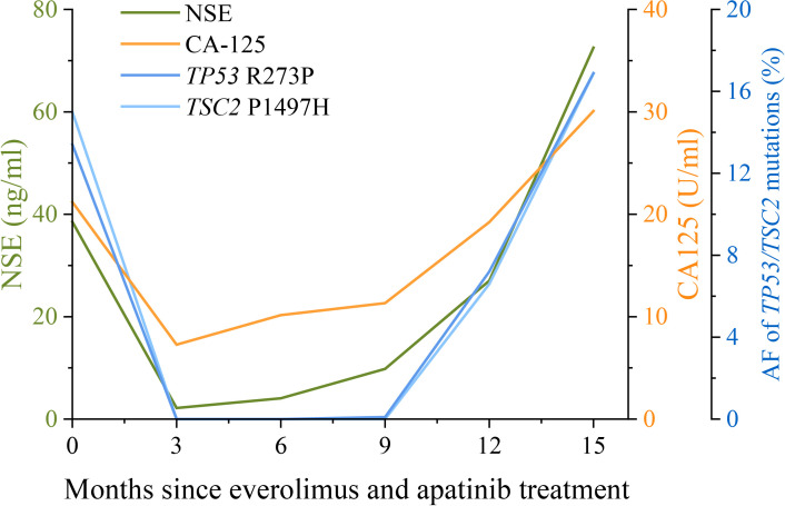
Perivascular epithelioid cell tumors (PEComas) are extremely rare mesenchymal neoplasms for which the uterus is the most common site. The prognosis of malignant PEComa is poor as it is characterized by resistance to classical chemotherapies. Both mTOR inhibitors and VEGFR inhibitors exhibited clinical utility in treating malignant PEComas, but the combination of these two regimens has rarely been reported. In the present case, a uterine PEComa patient developed lung and bone metastases after the failure of chemotherapies and derived benefit from the combination regimen of an mTOR inhibitor (everolimus) and a VEGFR inhibitor (apatinib), achieving a 15-month progression-free survival. Targeted NGS revealed TP53 and TSC2 mutations in the patient's primary uterine tumors and plasma ctDNA at disease progression. Plasma ctDNA clearance was consistent with a radiologic partial response determined by RECIST 1.1 and a reduction of neuron-specific enolase (NSE) and cancer antigen 125 (CA125) levels. Thus, we provided clinical evidence supporting the administration of combined therapy of mTOR and VEGFR inhibitors to metastatic uterine PEComa patients and highlighted the application of serial plasma ctDNA profiling for dynamic disease monitoring.

Keywords: TSC2; apatinib; everolimus; lung metastasis; uterine PEComa.


Conflict of interest statement

Authors EP, PY, TW, YM, and QO are employed by Nanjing Geneseeq Technology Inc. The remaining authors declare that the research was conducted in the absence of any commercial or financial relationships that could be construed as a potential conflict of interest.

Figures

Figure 1
Figure 1
Treatment history and clinical information of the presented case. (A) The medical history of the presented case is shown with information about treatment timeline, response evaluation, and sample collection timepoints. During the combination treatment with everolimus and apatinib, plasma ctDNA sequencing was performed every 3 months along with treatment response evaluation as indicated by the arrowheads. (B) H&E staining and immunohistochemical (IHC) examinations (×200) of the primary uterine PEComa which was negative for the SMA marker and positive for Melan-A (90%). The Ki67 index is 20%. (C) CT images of lung metastases during everolimus and apatinib treatment. Lesions are indicated by the red arrows. PR, partial response; SD, stable disease; PD, progressive disease; NGS, next-generation sequencing; PEComa, perivascular epithelioid cell tumors; ctDNA, circulating tumor DNA; FFPE, formalin-fixed, paraffin-embedded; H&E, hematoxylin and eosin; SMA, smooth muscle actin.
Figure 2
Figure 2
Changes in NSE and CA125 levels, and the allele frequencies (AFs) of TP53 and TSC2 mutations during apatinib and everolimus treatment. The levels of the lung cancer biomarkers NSE (neuron-specific enolase) and CA125 (cancer antigen 125) in serum examined every 3 months are shown by the green and orange lines, respectively. Plasma ctDNA sequencing was also performed every 3 months during apatinib and everolimus treatment. The AFs of TP53 R273P (blue) and TSC2 P1497H (light blue) mutations are shown by the dark blue and light blue lines, respectively. The units of NSE, CA125, and AF were indicated by the different y-axes.

Similar articles

Cited by

References

    1. Jo VY, Fletcher CD. WHO classification of soft tissue tumours: an update based on the 2013 (4th) edition. Pathology (2014) 46(2):95–104. doi: 10.1097/pat.0000000000000050 - DOI - PubMed
    1. Bennett JA, Braga AC, Pinto A, Van de Vijver K, Cornejo K, Pesci A, et al. . Uterine PEComas: A morphologic, immunohistochemical, and molecular analysis of 32 tumors. Am J Surg Pathol (2018) 42(10):1370–83. doi: 10.1097/PAS.0000000000001119 - DOI - PMC - PubMed
    1. Armah HB, Parwani AV. Perivascular epithelioid cell tumor. Arch Pathol Lab Med (2009) 133(4):648–54. doi: 10.5858/133.4.648 - DOI - PubMed
    1. Koshkin VS, Mir MC, Barata P, Gul A, Gupta R, Stephenson AJ, et al. . Randomized phase II trial of neoadjuvant everolimus in patients with high-risk localized prostate cancer. Investigational New Drugs (2019) 37(3):559–66. doi: 10.1007/s10637-019-00778-4 - DOI - PubMed
    1. Italiano A, Delcambre C, Hostein I, Cazeau AL, Marty M, Avril A, et al. . Treatment with the mTOR inhibitor temsirolimus in patients with malignant PEComa. Ann Oncol (2010) 21(5):1135–7. doi: 10.1093/annonc/mdq044 - DOI - PubMed

Publication types