

A protocol for rapid construction of senescent cells
- PMID: 35965600
- PMCID: PMC9372585
- DOI: 10.3389/fnint.2022.929788
A protocol for rapid construction of senescent cells
Abstract
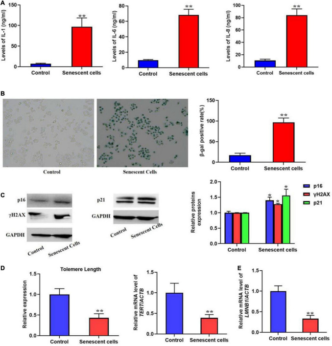
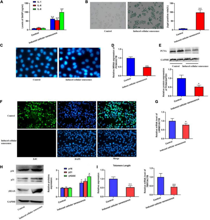
Aging may be the largest factor for a variety of chronic diseases that influence survival, independence, and wellbeing. Evidence suggests that aging could be thought of as the modifiable risk factor to delay or alleviate age-related conditions as a group by regulating essential aging mechanisms. One such mechanism is cellular senescence, which is a special form of most cells that are present as permanent cell cycle arrest, apoptosis resistance, expression of anti-proliferative molecules, acquisition of pro-inflammatory, senescence-associated secretory phenotype (SASP), and others. Most cells cultured in vitro or in vivo may undergo cellular senescence after accruing a set number of cell divisions or provoked by excessive endogenous and exogenous stress or damage. Senescent cells occur throughout life and play a vital role in various physiological and pathological processes such as embryogenesis, wound healing, host immunity, and tumor suppression. In contrast to the beneficial senescent processes, the accumulation of senescent also has deleterious effects. These non-proliferating cells lead to the decrease of regenerative potential or functions of tissues, inflammation, and other aging-associated diseases because of the change of tissue microenvironment with the acquisition of SASP. While it is understood that age-related diseases occur at the cellular level from the cellular senescence, the mechanisms of cellular senescence in age-related disease progression remain largely unknown. Simplified and rapid models such as in vitro models of the cellular senescence are critically needed to deconvolute mechanisms of age-related diseases. Here, we obtained replicative senescent L02 hepatocytes by culturing the cells for 20 weeks. Then, the conditioned medium containing supernatant from replicative senescent L02 hepatocytes was used to induce cellular senescence, which could rapidly induce hepatocytes into senescence. In addition, different methods were used to validate senescence, including senescence-associated β-galactosidase (SA-β-gal), the rate of DNA synthesis using 5-ethynyl-2'-deoxyuridine (EdU) incorporation assay, and senescence-related proteins. At last, we provide example results and discuss further applications of the protocol.
Keywords: SASP; aging; biomarker; cellular senescence; senescent cell models.
Copyright © 2022 Yu, Quan, Chen, Yang, Huang, Yang and Zhang.
Conflict of interest statement
The authors declare that the research was conducted in the absence of any commercial or financial relationships that could be construed as a potential conflict of interest.
Figures
References
LinkOut - more resources
Full Text Sources