

Central Odontogenic Fibroma Accompanied by a Central Giant Cell Granuloma-Like Lesion: Report of a Case and Review of Literature
- PMID: 35965713
- PMCID: PMC9355841
- DOI: 10.18502/fid.v18i44.8340
Central Odontogenic Fibroma Accompanied by a Central Giant Cell Granuloma-Like Lesion: Report of a Case and Review of Literature
Abstract
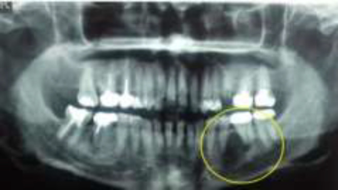
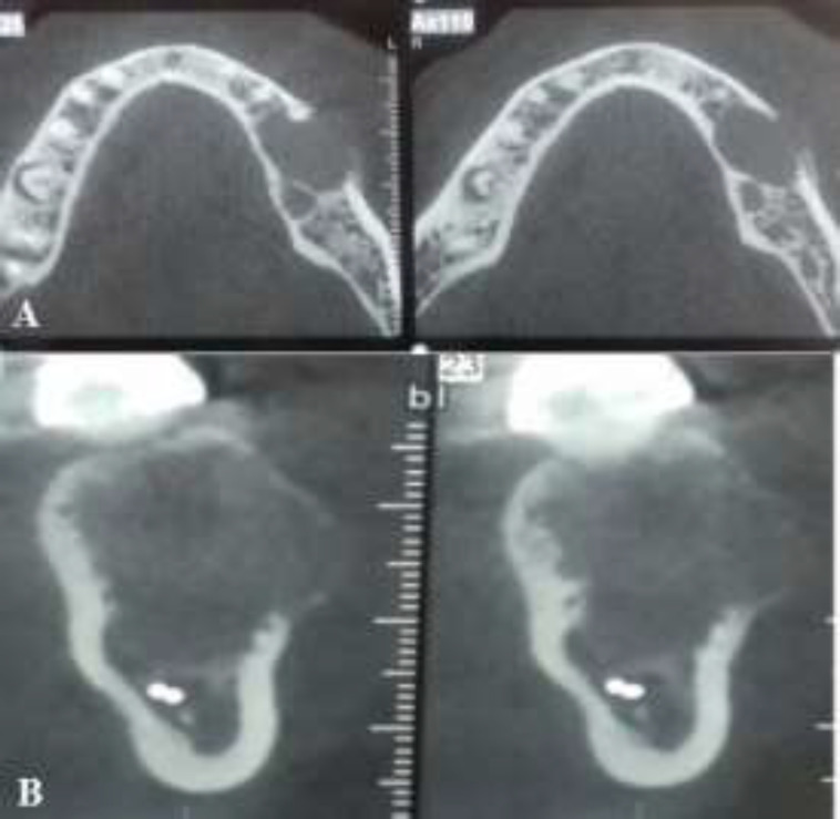
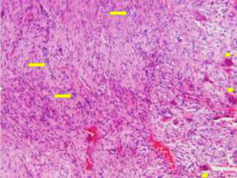
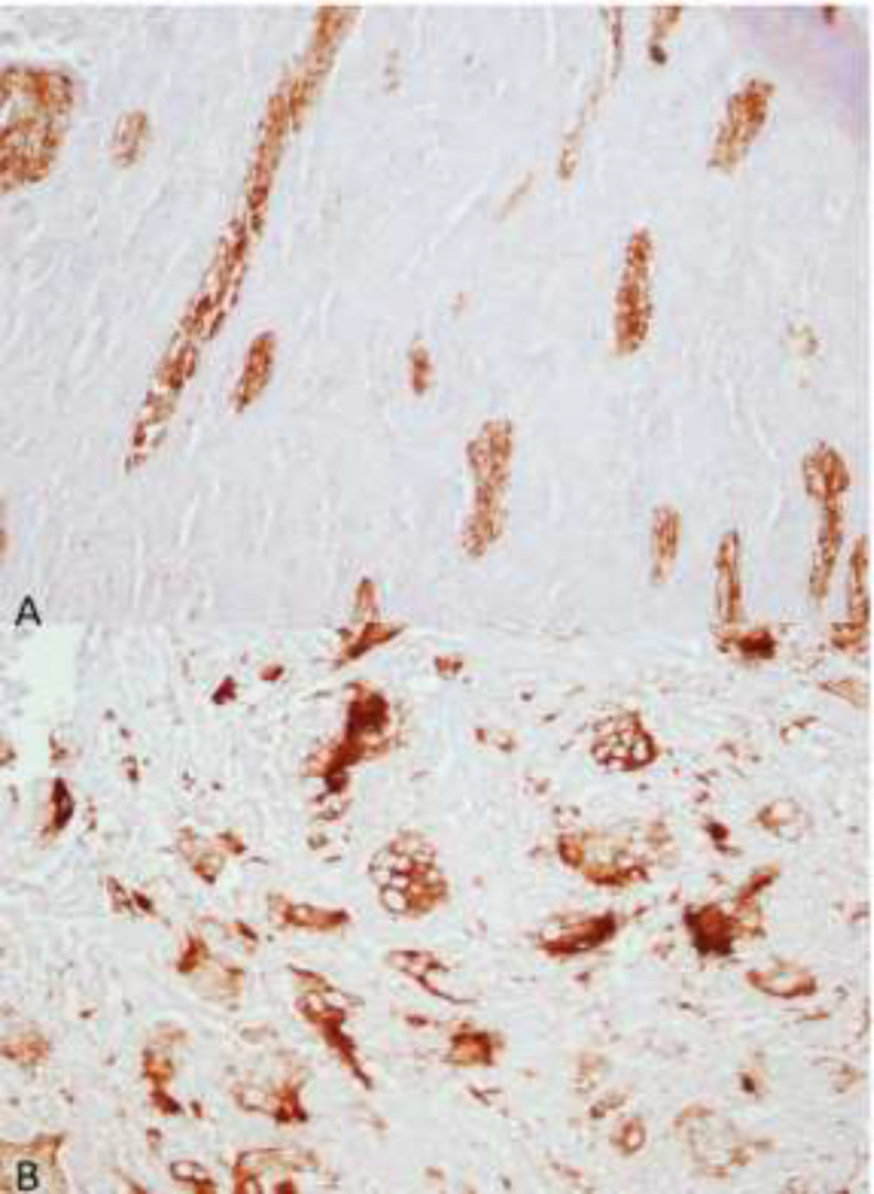
Central giant cell granuloma (CGCG) is a benign non-neoplastic intraosseous lesion mainly found in the anterior mandible. It is characterized by multinucleated giant cells, representing osteoclasts or macrophages. Central odontogenic fibroma (COF) is an uncommon benign lesion of the jaws. It originates from the odontogenic ectomesenchyme. In rare cases, COF may accompany a CGCG. To date, 49 cases of COF accompanied by CGCG-like lesions have been reported in the literature. In this paper, we present another case of COF-CGCG in a 46-year-old female. The lesion was located in the posterior mandible. Excisional biopsy was carried out, and histopathological analysis revealed multinucleated giant cells with numerous strands of odontogenic epithelium. A literature review of previously reported cases was also performed.
Keywords: Fibroma; Granuloma, Giant Cell; Odontogenic Tumors.
Copyright © 2021 The Authors. Published by Tehran University of Medical Sciences.
Figures
Similar articles
-
Central odontogenic fibroma: retrospective study of six cases with variable histopathologic features using 2022 WHO classification.BMC Oral Health. 2024 Oct 26;24(1):1297. doi: 10.1186/s12903-024-05085-w. BMC Oral Health. 2024. PMID: 39462411 Free PMC article.
-
Hybrid Central Odontogenic Fibroma with Giant Cell Granuloma like Lesion: A Report of Three Additional Cases and Review of the Literature.Head Neck Pathol. 2018 Jun;12(2):166-174. doi: 10.1007/s12105-017-0845-7. Epub 2017 Aug 7. Head Neck Pathol. 2018. PMID: 28785965 Free PMC article. Review.
-
So-called hybrid central odontogenic fibroma/central giant cell lesion of the jaws. A report on seven additional cases, including an example in a patient with cherubism, and hypotheses on the pathogenesis.Head Neck Pathol. 2008 Dec;2(4):333-8. doi: 10.1007/s12105-008-0076-z. Epub 2008 Aug 23. Head Neck Pathol. 2008. PMID: 20614305 Free PMC article.
-
Hybrid central odontogenic fibroma with giant cell granuloma-like component: case report and review of literature.Head Neck Pathol. 2008 Sep;2(3):222-6. doi: 10.1007/s12105-008-0063-4. Epub 2008 Jun 10. Head Neck Pathol. 2008. PMID: 20614319 Free PMC article. Review.
-
Rare hybrid tumor of odontogenic fibromyxoma and central giant cell granuloma in maxilla: First reported case.Indian J Dent Res. 2023 Jul-Sep;34(3):332-334. doi: 10.4103/ijdr.ijdr_349_22. Indian J Dent Res. 2023. PMID: 38197359
Cited by
-
Fibrous dysplasia associated with peripheral giant cell granoluma in maxilla in a young patient, a case report of rare hybrid lesion.Rare Tumors. 2023 Apr 24;15:20363613231165883. doi: 10.1177/20363613231165883. eCollection 2023. Rare Tumors. 2023. PMID: 37124839 Free PMC article.
-
Central odontogenic fibroma: retrospective study of six cases with variable histopathologic features using 2022 WHO classification.BMC Oral Health. 2024 Oct 26;24(1):1297. doi: 10.1186/s12903-024-05085-w. BMC Oral Health. 2024. PMID: 39462411 Free PMC article.
References
-
- Kruse-Losler B, Diallo R, Gaertner C, Mischke KL, Joos U, Kleinheinz J. Central giant cell granuloma of the jaws: a clinical, radiologic, and histopathologic study of 26 cases. Oral Surg Oral Med Oral Pathol Oral Radiol Endod. 2006 Mar;101(3):346–54. - PubMed
-
- de Lange J, van den Akker HP, van den Berg H. Central giant cell granuloma of the jaw: a review of the literature with emphasis on therapy options. Oral Surg Oral Med Oral Pathol Oral Radiol Endod. 2007 Nov;104(5):603–15. - PubMed
Publication types
LinkOut - more resources
Full Text Sources