. 2022 Jul;10(14):776.
doi: 10.21037/atm-22-2770.

Interleukin-35 attenuates blood-brain barrier dysfunction caused by cerebral ischemia-reperfusion injury through inhibiting brain endothelial cell injury

Affiliations

Interleukin-35 attenuates blood-brain barrier dysfunction caused by cerebral ischemia-reperfusion injury through inhibiting brain endothelial cell injury

Lei Qian et al. Ann Transl Med. 2022 Jul.

Abstract

Background: Interleukin-35 (IL-35), an anti-inflammatory and antioxidant cytokine, plays a potent immunosuppressive role in various diseases. However, the effects of IL-35 on blood-brain barrier (BBB) dysfunction in ischemic stroke are not well characterized.

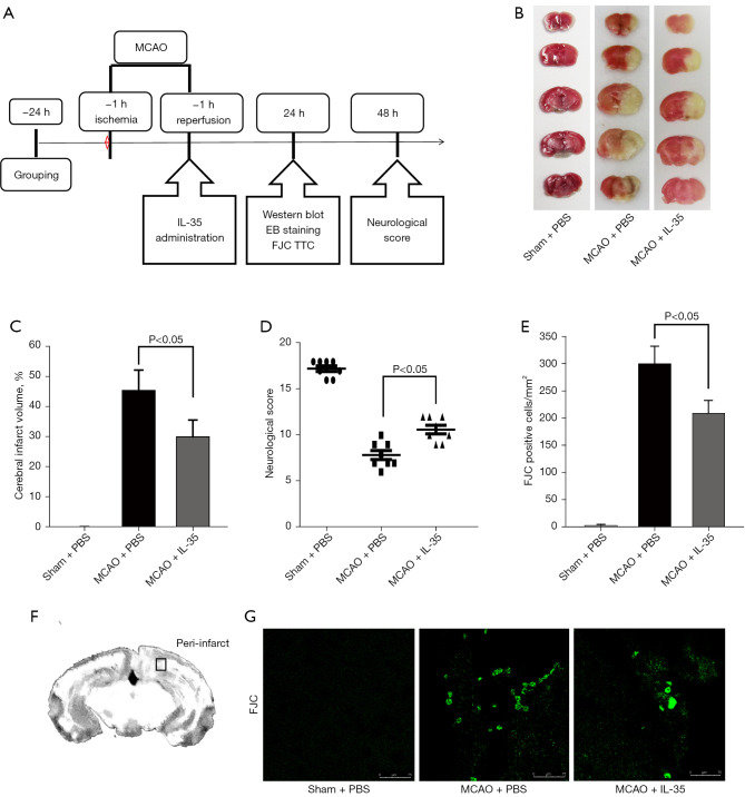
Methods: A total of 150 male C57BL/6 mice (aged 6-8 weeks and weighing 20-25 g) were used in this study. The protective effects of IL-35 against BBB dysfunction were examined using a mouse model of middle cerebral artery occlusion (MCAO) and an in vitro model of oxygen-glucose deprivation/reoxygenation (OGD/R) injury in mouse brain endothelial cells (bEnd.3).

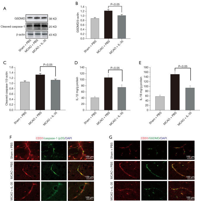
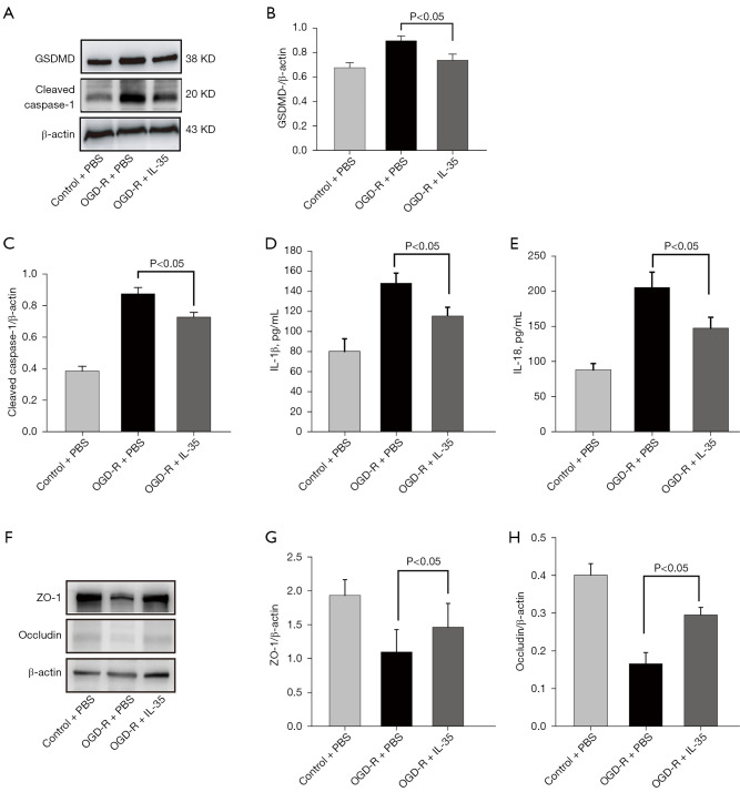
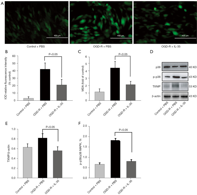
Results: Intracerebroventricular administration of IL-35 (10 µg/g) was found to reduce cerebral edema and Evans blue (EB) leakage, and increase the expression of tight junction (TJ) proteins, thereby attenuating MCAO-induced neurological deficit in mice. Moreover, IL-35 (20 ng/mL) treatment upregulated the expression of TJ proteins in OGD/R-induced bEnd.3 cells. IL-35 also markedly suppressed the expression of caspase-1, IL-1β, and gasdermin D (GSDMD) in vivo and in vitro. In addition, IL-35 decreased the generation of reactive oxygen species (ROS) and inhibited the expression of thioredoxin-interacting protein (TXNIP) in OGD/R-induced bEnd.3 cells.

Conclusions: These results indicated that IL-35 exerts a protective effect on the BBB by targeting the ROS/TXNIP/caspase-1 pathway in cerebral ischemia-reperfusion (I/R) injury.

Keywords: Ischemic stroke; blood-brain barrier (BBB); caspase-1; interleukin-35 (IL-35).


Conflict of interest statement

Conflicts of Interest: All authors have completed the ICMJE uniform disclosure form (available at https://atm.amegroups.com/article/view/10.21037/atm-22-2770/coif). The authors have no conflicts of interest to declare.

Figures

Figure 1
Figure 1
IL-35 treatment ameliorated I/R-induced cerebral infarction and neurological deficit. (A) Experimental protocol in mice subjected to MCAO. IL-35 (10 µg/g) was administered at 1 hour and 24 hours after reperfusion by intracerebroventricular injection. (B,C) Representative images and statistical analysis of the infarct areas. The infarct volume was quantified using TTC staining at 24 hours after ischemia (n=6 per group). (D) Neurological scores at 48 hours after reperfusion according to the Garcia scoring method (n=8 per group). (E-G) Representative images and statistical analysis of neuronal degeneration. FJC staining was performed 24 hours after I/R (scale bar: 75 µm). Data are presented as mean ± SD (n=6 per group). MCAO, middle cerebral artery occlusion; IL, interleukin; EB, Evans blue; FJC, fluoro-Jade C; TTC, 2,3,5-triphenyltetrazolium chloride; PBS, phosphate buffered saline; I/R, ischemia/reperfusion; SD, standard deviation.
Figure 2
Figure 2
IL-35 suppressed I/R-induced BBB dysfunction. MCAO mice were administered IL-35 (10 µg/g) or PBS until sacrificed. (A) Representative images and statistical analysis of EB leakage analysis at 24 hours after reperfusion. (B) Brain water content analysis of the injured hemisphere at 24 hours after reperfusion. (C-E) Western blot images and analysis of ZO-1 and occludin expressions in the peri-ischemic region at 24 hours after reperfusion. Data presented as mean ± SD (n=6 per group). (F,G) Immunofluorescence staining of ZO-1 and occludin (green) expression in vascular endothelial cells (CD31, red) in MCAO mice brain coronal sections (scale bar: 100 µm; n=6 per group). MCAO, middle cerebral artery occlusion; IL, interleukin; PBS, phosphate buffered saline; ZO-1, zonula occludens-1; I/R, ischemia/reperfusion; BBB, blood-brain barrier; EB, Evans blue; SD, standard deviation.
Figure 3
Figure 3
IL-35 suppressed I/R-induced brain tissue injury in the ischemic cortex. MCAO mice were administered IL-35 (10 µg/g) or PBS and sacrificed at 24 hours after reperfusion. (A-C) Western blot images and analysis of cleaved caspase-1 and GSDMD expressions in the peri-ischemic region. Error bars represent mean ± SD; n=6 per group. (D,E) Results of ELISA showing the concentrations of IL-18 and IL-1β in the cortical tissues. Error bars represent mean ± SD; n=6 per group. (F,G) Immunofluorescence staining of cleaved caspase-1 and GSDMD (green) expression in vascular endothelial cells (CD31, red) in the peri-ischemic regions (scale bar: 100 µm; n=6 per group). GSDMD, gasdermin D; PBS, phosphate buffered saline; MCAO, middle cerebral artery occlusion; IL, interleukin; I/R, ischemia/reperfusion; SD, standard deviation; ELISA, enzyme-linked immunosorbent assay.
Figure 4
Figure 4
IL-35 protected OGD/R-induced endothelial cell injury. bEnd.3 cells were pretreated with or without IL-35 and then subjected to 6 hours of OGD and 18 hours reoxygenation. (A-C) Western blot images and analysis of cleaved caspase-1 and GSDMD expressions in bEnd.3 cells. (D,E) Results of ELISA showing the levels of IL-18 and IL-1β in the culture supernatant. (F-H) Results of Western blot assay showing the protein expressions of ZO-1 and occludin in bEnd.3 cells. Error bars represent mean ± SD of at least three independent experiments. GSDMD, gasdermin D; PBS, phosphate buffered saline; OGD-R, oxygen-glucose deprivation and reperfusion; IL, interleukin; ZO-1, zonula occludens-1; bEnd.3, brain endothelial cells; ELISA, enzyme-linked immunosorbent assay; SD, standard deviation.
Figure 5
Figure 5
IL-35 suppressed OGD/R-induced ROS generation in endothelial cells. The effects of IL-35 on ROS generation and the MAPK pathway in bEnd.3 cells after 6 hours of OGD and 18 hours reoxygenation. (A) Intracellular ROS was viewed with fluorescence microscopy (scale bar: 400 µm). (B) The resulting fluorescence intensity was quantified using Image J software. Error bars represent mean ± SD of at least three independent experiments. (C) The concentration of MDA in cell lysates. (D-F) Representative Western blot bands and quantitative analysis of the ratio of p-p38/p38 and TXNIP. The band intensities were measured using scanning densitometry. Protein expressions were normalized to β-actin (n=3). Data are expressed as mean ± SD. PBS, phosphate buffered saline; OGD-R, oxygen-glucose deprivation and reperfusion; IL, interleukin; IOD, integrated option density; MDA, malondialdehyde; TXNIP, thioredoxin-interacting protein; ROS, reactive oxygen species; MAPK, mitogen-activated proliferation protein kinase; SD, standard deviation.

Similar articles

Cited by

References

    1. Feske SK. Ischemic Stroke. Am J Med 2021;134:1457-64. 10.1016/j.amjmed.2021.07.027 - DOI - PubMed
    1. Paul S, Candelario-Jalil E. Emerging neuroprotective strategies for the treatment of ischemic stroke: An overview of clinical and preclinical studies. Exp Neurol 2021;335:113518. 10.1016/j.expneurol.2020.113518 - DOI - PMC - PubMed
    1. Sarvari S, Moakedi F, Hone E, et al. Mechanisms in blood-brain barrier opening and metabolism-challenged cerebrovascular ischemia with emphasis on ischemic stroke. Metab Brain Dis 2020;35:851-68. 10.1007/s11011-020-00573-8 - DOI - PMC - PubMed
    1. Maida CD, Norrito RL, Daidone M, et al. Neuroinflammatory Mechanisms in Ischemic Stroke: Focus on Cardioembolic Stroke, Background, and Therapeutic Approaches. Int J Mol Sci 2020;21:6454. 10.3390/ijms21186454 - DOI - PMC - PubMed
    1. Yawoot N, Govitrapong P, Tocharus C, et al. Ischemic stroke, obesity, and the anti-inflammatory role of melatonin. Biofactors 2021;47:41-58. 10.1002/biof.1690 - DOI - PubMed