. 2022 Jul;10(14):768.
doi: 10.21037/atm-22-3131.

The efficacy and safety of blunt impingement followed by a sharp recanalization technique in hemodialysis patients with refractory central vein occlusion: a single-center experience

Affiliations

The efficacy and safety of blunt impingement followed by a sharp recanalization technique in hemodialysis patients with refractory central vein occlusion: a single-center experience

Ji-Bo Sun et al. Ann Transl Med. 2022 Jul.

Abstract

Background: Central vein occlusion (CVO) is a serious problem in hemodialysis patients. There is an unsatisfactory result for refractory CVO by sharp recanalization alone. This study evaluated the efficacy and safety of blunt impingement followed by sharp recanalization for the treatment of CVO in hemodialysis patients.

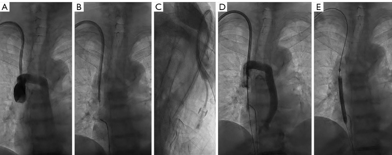
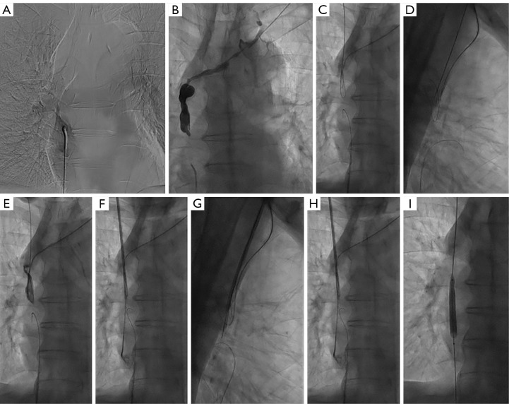
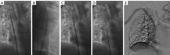
Methods: This study retrospectively examined hemodialysis patients with CVO who failed to recanalize using standard guidewire and catheter techniques in our department. In the first instance, all CVOs were recanalized using blunt impingement techniques, including a 6-Fr long sheath (Cook Incorporated, Bloomington, IN USA) and an 8-Fr sheath of Rosch-Uchida Transjugular Liver Access Set (RUPS-100; Cook Incorporated, Bloomington, IN, USA). If this was not successful, sharp recanalization devices were applied, including the stiff tip of a guidewire (Terumo, Tokyo, Japan), the RUPS-100, and the percutaneous transhepatic cholangial drainage (PTCD) needle (Cook Incorporated, USA). All patients were followed up at least 4 months postoperatively. The technical success rate, arteriovenous access patency rates, and operation-related complications were analyzed.

Results: The procedural success rate was 100.0% (30 of 30). Thirty patients with CVO underwent blunt impingement with a technique success rate of 70.0% (21 of 30), and 9 patients received sharp recanalization after failed blunt impingement, with a technique success rate of 100.0% (9 of 9). The primary patency rates at 6 and 12 months postoperatively were 86.7% and 53.3%, respectively. The primary assisted patency rates were 93.3% and 63.3%, and the secondary patency rates were 93.3% and 70.0% at 6 and 12 months, respectively. One major procedure-related complication was detected, namely, a small injury of the superior vena cava (SVC) wall in a patient receiving recanalization via the stiff end of a guidewire, but this did not require further treatment.

Conclusions: It is potentially effective and safe for interventionalists to use blunt impingement followed by sharp recanalization techniques to treat chronic CVO that is refractory to traversal using traditional catheter and guidewire techniques.

Keywords: Blunt impingement; hemodialysis; refractory central venous occlusion; sharp recanalization.


Conflict of interest statement

Conflicts of Interest: All authors have completed the ICMJE uniform disclosure form (available at https://atm.amegroups.com/article/view/10.21037/atm-22-3131/coif). The authors have no conflicts of interest to declare.

Figures

Figure 1
Figure 1
Procedure of the blunt impingement technique for recanalization of the occlusion of the RSV and the RBV. (A) Venography from the arteriovenous fistula of the right upper extremity showing the distal end of subclavian venous occlusion. (B) A 6-Fr introducer catheter was introduced into the lesion through the arteriovenous fistula. (C) A 4-Fr angiographic catheter was used to impinge the occluded segment of the RSV until it successfully crosses the lesion. (D) The 4-Fr angiographic catheter was used to repeatedly impinge the distal end of the RBV occlusion, with assistance and sufficient support from a 6-Fr long sheath, without success. (E) A 9-Fr long sheath was then introduced into the distal end of the SVC to act as a target for the 4-Fr angiographic catheter blunt impingement. (F) The distal end of the RBV occlusion was impinged again using a 4-Fr angiographic catheter and the SVC was reached successfully. (G) A guidewire from the femoral vein approach was snared from the SV. (H) A venogram showing the LSV was still narrow after balloon dilation. (I) An angiography indicted that the lesion was still narrow after the implantation of the 10-mm covered stent (W. L. Gore & Associates, USA). (J) A repeat venography revealed that complete restoration of patency was achieved after placement of the 10-mm bare stent (Cordis Corporation, USA). RSV, right subclavian vein; RBV, right brachiocephalic vein; SVC, superior vena cava; SV, subclavian vein; LSV, left subclavian vein.
Figure 2
Figure 2
The procedure for the blunt impingement technique for the LBV occlusion. (A) An angiography showing complete occlusion of the LBV. (B) With the support of a 6-long sheath, a 4-Fr angiographic catheter was used to impinge the lesion. (C) Balloon angioplasty of the occlusive LBV. (D) Postangioplasty venography revealed complete restoration of patency of the LBV. LBV, left brachiocephalic vein.
Figure 3
Figure 3
The stiff tip of the guidewire was used to recanalize the SVC occlusion. (A) A venogram showing the SVC occlusion. (B,C) The stiff tip of a hydrophilic guidewire crossed the SVC occlusion. (D) A second angiogram showing that a small amount of contrast was able to enter the right atrium through the residual slit in the occluded segment, and the hydrophilic guidewire passed the lesion successfully. (E) The lesion recovered well after balloon dilation. SVC, superior vena cava.
Figure 4
Figure 4
The RUPS-100 recanalized the SVC occlusion. (A,B) The venography revealed complete occlusion of the SVC. (C,D) The SVC was punctured using a PTCD needle from below the lateral head of the sternocleidomastoid muscle via the right neck. (E) The PTCD needle was placed at the distal end of the SVC. (F,G) A stiff guidewire was introduced into the RUPS-100 metal sheath from the right neck. (H) A venogram showing a 4-Fr catheter located in the right atrium after successful sharp recanalization. (I) A 6-mm balloon dilation. RUPS, Rosch-Uchida Transjugular Liver Access Set; SVC, superior vena cava; PTCD, percutaneous transhepatic cholangial drainage.
Figure 5
Figure 5
The procedure for PTCD needle puncture of the occlusion of the RBV. (A) and (B) A venogram showing the PTCD needle directly punctured the distal end of the RBV using a snare as a target. (C) The PTCD sheath was placed in the SVC. (D) The second snare grasped a guidewire from the right femoral vein access. (E) The lesion recovered well after balloon dilation and stenting. PTCD, percutaneous transhepatic cholangial drainage; RBV, right brachiocephalic vein; SVC, superior vena cava.
Figure 6
Figure 6
Kaplan-Meier curves of primary, primary assisted, and secondary patency rates post-operation.

References

    1. Toomay S, Rectenwald J, Vazquez MA. How Can the Complications of Central Vein Catheters Be Reduced?: Central Venous Stenosis in Hemodialysis Patients. Semin Dial 2016;29:201-3. 10.1111/sdi.12478 - DOI - PubMed
    1. Lumsden AB, MacDonald MJ, Isiklar H, et al. Central venous stenosis in the hemodialysis patient: incidence and efficacy of endovascular treatment. Cardiovasc Surg 1997;5:504-9. 10.1016/S0967-2109(97)00043-4 - DOI - PubMed
    1. Anaya-Ayala JE, Smolock CJ, Colvard BD, et al. Efficacy of covered stent placement for central venous occlusive disease in hemodialysis patients. J Vasc Surg 2011;54:754-9. 10.1016/j.jvs.2011.03.260 - DOI - PubMed
    1. Lok CE, Huber TS, Lee T, et al. KDOQI Clinical Practice Guideline for Vascular Access: 2019 Update. Am J Kidney Dis 2020;75:S1-S164. 10.1053/j.ajkd.2019.12.001 - DOI - PubMed
    1. Kundu S. Review of central venous disease in hemodialysis patients. J Vasc Interv Radiol 2010;21:963-8. 10.1016/j.jvir.2010.01.044 - DOI - PubMed