

Risk factors for bone cement displacement after percutaneous vertebral augmentation for osteoporotic vertebral compression fractures
- PMID: 35965863
- PMCID: PMC9366098
- DOI: 10.3389/fsurg.2022.947212
Risk factors for bone cement displacement after percutaneous vertebral augmentation for osteoporotic vertebral compression fractures
Abstract
Purpose: To explore the risk factors of bone cement displacement after percutaneous vertebral augmentation (PVA) in patients with osteoporotic vertebral compression fracture (OVCF).
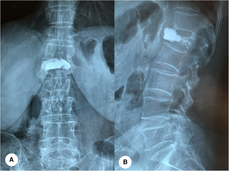
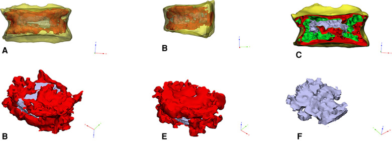
Methods: We retrospectively reviewed the records of 1,538 patients with OVCF treated with percutaneous vertebroplasty (PVP) or percutaneous vertebroplasty (PKP) from January 2016 to June 2021. Patients were divided into bone cement displacement group (n = 78) and bone cement non-displacement group (n = 1,460) according to the radiographic images. Possible risk factors for bone cement displacement were noted, including age, gender, body mass index (BMI), bone mineral density (BMD), underlying disease, number of fractured vertebrae, involved vertebral segment, surgical method, surgical approach, vertebral height, Cobb angle, cement leakage, the viscosity of bone cement, bone cement diffuse ratio, degree of bone cement interweaving, sagittal bone cement placement, targeted location of bone cement, the distance between the bone cement and the upper and lower endplates, the time of wearing brace and postoperative osteoporosis treatment. Risk factors were identified with univariate and multivariate logistic regressions and the discrimination ability of the predictive indicators was evaluated using area under the curve (AUC) of the receiver operating characteristic (ROC).
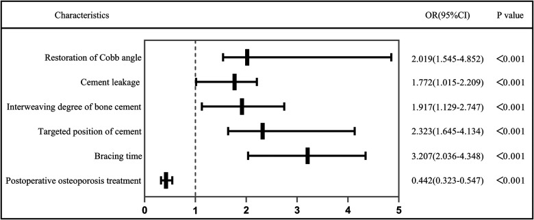
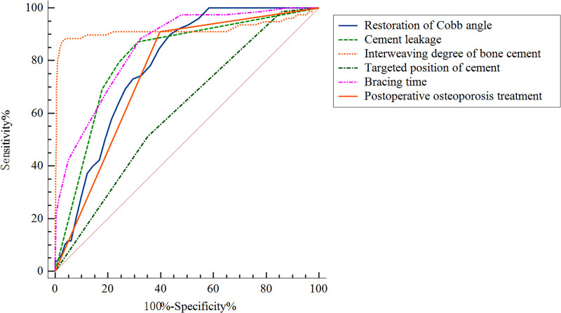
Results: In multivariate regression, independent risk factors for bone cement displacement included: high restoration of Cobb angle (OR = 2.019, 95%[CI] 1.545-4.852, P < 0.001), cement leakage (anterior edge) (OR = 1.727, 95%[CI] 1.05-2.20, P < 0.001), small degree of bone cement interweaving (OR = 1.917, 95%[CI] 1.129-2.747, P < 0.001), non-targeted location of bone cement (OR = 2.323, 95%[CI] 1.645-4.134, P < 0.001), short duration of brace wearing (OR = 3.207, 95%[CI] 2.036-4.348, P < 0.001) and postoperative osteoporosis treatment (OR = 0.422, 95% CI = 0.323-0.547, P < 0.001). The AUCs for the high restoration of Cobb angle, cement leakage (anterior edge), small degree of bone cement interweaving, non-targeted location of bone cement, short duration of brace wearing and non-postoperative osteoporosis treatment were 0.784 (95% CI, 0.747-0.821), 0.811 (95% CI 0.764-0.859), 0.917 (95%CI 0.864-0.970), 0.610 (95%CI 0.552-0.669), 0.854 (95%CI 0.816-0.892) and 0.756 (95% CI, 0.712-0.800), respectively.
Conclusion: High restoration of Cobb angle, cement leakage (anterior edge), small degree of bone cement interweaving, non-targeted location of bone cement, short duration of brace wearing and non-postoperative osteoporosis treatment were the independent risk factors of bone cement displacement after PVA.
Keywords: bone cement displacement; complication; osteoporotic vertebral compression fracture (OVCF); percutaneous vertebral augmentation; risk factors.
© 2022 Gao, Du, Gao, Hao, Hui, He and Yan.
Conflict of interest statement
The authors declare that the research was conducted in the absence of any commercial or financial relationships that could be construed as a potential conflict of interest.
Figures
Similar articles
-
Predictive Factors for Bone Cement Displacement following Percutaneous Vertebral Augmentation in Kümmell's Disease.J Clin Med. 2022 Dec 16;11(24):7479. doi: 10.3390/jcm11247479. J Clin Med. 2022. PMID: 36556095 Free PMC article.
-
Logistic regression analysis on risk factors of augmented vertebra recompression after percutaneous vertebral augmentation.J Orthop Surg Res. 2021 Jun 11;16(1):374. doi: 10.1186/s13018-021-02480-9. J Orthop Surg Res. 2021. PMID: 34116683 Free PMC article.
-
[Correlation analysis of cement leakage with volume ratio of intravertebral bone cement to vertebral body and vertebral body wall incompetence in percutaneous vertebroplasty for osteoporotic vertebral compression fractures].Zhongguo Xiu Fu Chong Jian Wai Ke Za Zhi. 2014 Nov;28(11):1358-63. Zhongguo Xiu Fu Chong Jian Wai Ke Za Zhi. 2014. PMID: 25639050 Chinese.
-
Comparison of Percutaneous Vertebroplasty and Balloon Kyphoplasty for the Treatment of Single Level Vertebral Compression Fractures: A Meta-analysis of the Literature.Pain Physician. 2015 May-Jun;18(3):209-22. Pain Physician. 2015. PMID: 26000665 Review.
-
Lumbar posterior group muscle degeneration: Influencing factors of adjacent vertebral body re-fracture after percutaneous vertebroplasty.Front Med (Lausanne). 2023 Apr 17;9:1078403. doi: 10.3389/fmed.2022.1078403. eCollection 2022. Front Med (Lausanne). 2023. PMID: 37138584 Free PMC article. Review.
Cited by
-
Risk factors for low back pain following percutaneous vertebroplasty in patients with osteoporotic vertebral compression fracture.Am J Transl Res. 2024 Aug 15;16(8):3778-3786. doi: 10.62347/SKKU1066. eCollection 2024. Am J Transl Res. 2024. PMID: 39262739 Free PMC article.
-
Clinical efficacy and biomechanical analysis of a novel hollow pedicle screw combined with kyphoplasty for the treatment of Kümmell disease.JOR Spine. 2024 Dec 6;7(4):e70017. doi: 10.1002/jsp2.70017. eCollection 2024 Dec. JOR Spine. 2024. PMID: 39649796 Free PMC article.
-
Evaluation and analysis of risk factors for adverse events of the fractured vertebra post-percutaneous kyphoplasty: a retrospective cohort study using multiple machine learning models.J Orthop Surg Res. 2024 Sep 18;19(1):575. doi: 10.1186/s13018-024-05062-7. J Orthop Surg Res. 2024. PMID: 39289697 Free PMC article.
-
Construction and clinical validation of risk model for predicting bone cement leakage after the surgical management of spinal metastases.Am J Cancer Res. 2024 Oct 15;14(10):4841-4854. doi: 10.62347/JAIR5009. eCollection 2024. Am J Cancer Res. 2024. PMID: 39553224 Free PMC article.
-
Comparison of percutaneous vertebroplasty and percutaneous vertebroplasty combined with pediculoplasty for Kümmell's disease: a retrospective observational study.J Orthop Surg Res. 2023 Jun 29;18(1):471. doi: 10.1186/s13018-023-03957-5. J Orthop Surg Res. 2023. PMID: 37386585 Free PMC article.
References
-
- Beall DP, Olan WJ, Kakad P, Li Q, Hornberger J. Economic analysis of kiva Vcf treatment system compared to balloon kyphoplasty using randomized kiva safety and effectiveness trial (kast) data. Pain Physician. (2015) 18(3):E299–E306. - PubMed
LinkOut - more resources
Full Text Sources
Miscellaneous