

Imprint Cytology Can Be a Better Option for Diagnosis of Mammary Paget's Disease!!!: A Case Report with Review
- PMID: 35965905
- PMCID: PMC9364448
- DOI: 10.4103/JCAS.JCAS_145_21
Imprint Cytology Can Be a Better Option for Diagnosis of Mammary Paget's Disease!!!: A Case Report with Review
Abstract
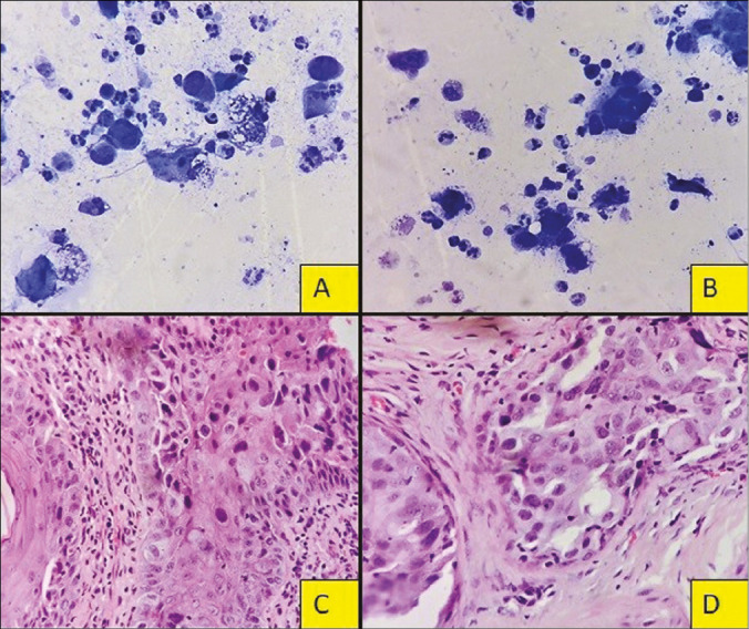
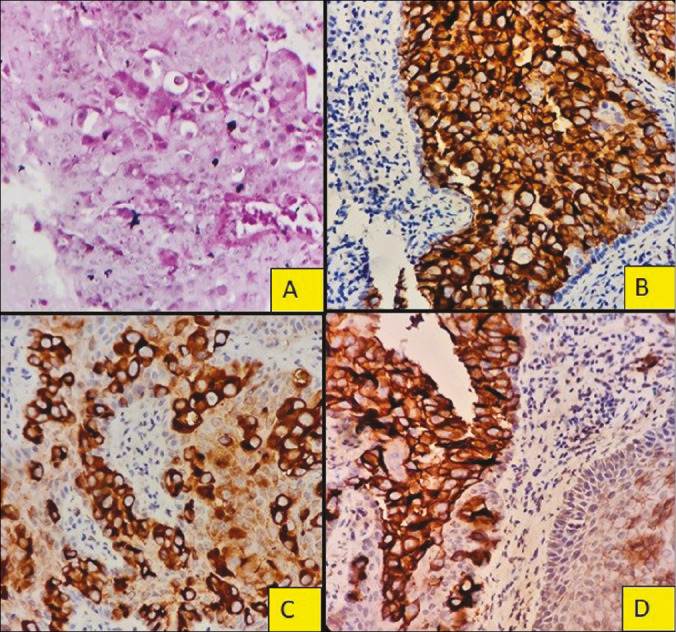
Mammary Paget's disease (MPD) is a rare form of pruritic eczematous skin lesion involving the nipple areola complex usually associated with an underlying in-situ or invasive carcinoma of breast, accounting ≤ 4% of overall breast carcinoma. The patient may present with nipple discharge, eczema, plaque, or nipple destruction with or without a lump which is resistant to common remedies. Diagnosis of MPD is usually accomplished by a punch biopsy, but imprint cytology is found to be an effective modern technique yet less explored. Cytological diagnosis is advantageous over surgical excisional biopsy; advantages being easy practicability, cost-effective, quick non-invasive, and above all can lead the physician for breast-preserving surgeries in selective cases rather than more aggressive standard mastectomies. We report one such case of MPD in a 52-year-old female diagnosed on imprint cytology with the aim to emphasize that imprint cytology can be a better option to improve the treatment protocol.
Keywords: Breast-conserving surgery; imprint cytology; mammary Paget.
Copyright: © 2022 Journal of Cutaneous and Aesthetic Surgery.
Conflict of interest statement
There are no conflicts of interest.
Figures
Similar articles
-
Paget's disease of the nipple diagnosed on cytology: a case report.Indian J Pathol Microbiol. 2004 Apr;47(2):246-8. Indian J Pathol Microbiol. 2004. PMID: 16295486
-
Mammary Paget's Disease Mimicking Benign and Malignant Dermatological Conditions: Clinical Challenges and Diagnostic Considerations.Cureus. 2024 Jul 25;16(7):e65378. doi: 10.7759/cureus.65378. eCollection 2024 Jul. Cureus. 2024. PMID: 39188449 Free PMC article. Review.
-
Paget's disease of the breast.Cancer Treat Rev. 2001 Feb;27(1):9-18. doi: 10.1053/ctrv.2000.0203. Cancer Treat Rev. 2001. PMID: 11237774 Review.
-
Pigmented Paget's disease of nipple: A diagnostic challenge on cytology.J Cytol. 2013 Jan;30(1):68-70. doi: 10.4103/0970-9371.107521. J Cytol. 2013. PMID: 23661948 Free PMC article.
-
Mammary Paget's disease without underlying malignancy of the breast.Yeungnam Univ J Med. 2018 Jun;35(1):99-103. doi: 10.12701/yujm.2018.35.1.99. Epub 2018 Jun 30. Yeungnam Univ J Med. 2018. PMID: 31620578 Free PMC article.
References
-
- Valdes EK, Feldman SM. Paget’s disease of the breast. Breast J. 2006;12:83. - PubMed
-
- Dixon AR, Galea MH, Ellis IO, Elston CW, Blamey RW. Paget’s disease of the nipple. Br J Surg. 1991;78:722–3. - PubMed
-
- Paget J. On disease of the mammary areola preceding cancer of the mammary gland. St Bartholomew’s Hosp Rep. 1874;10:87–9.
-
- Sakorafas GH, Blanchard K, Sarr MG, Farley DR. Paget’s disease of the breast. Cancer Treat Rev. 2001;27:9–18. - PubMed
Publication types
LinkOut - more resources
Full Text Sources