

Plasmacytoid Dendritic Cell Marker (CD123) Expression in Scarring and Non-scarring Alopecia
- PMID: 35965907
- PMCID: PMC9364453
- DOI: 10.4103/JCAS.JCAS_126_19
Plasmacytoid Dendritic Cell Marker (CD123) Expression in Scarring and Non-scarring Alopecia
Abstract
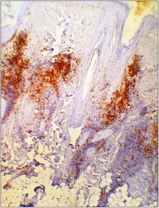
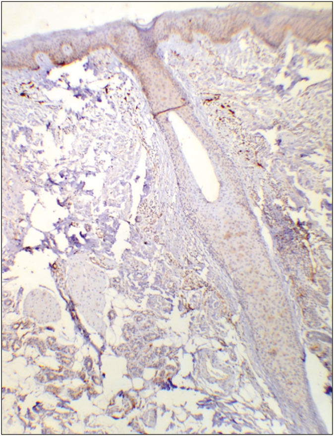
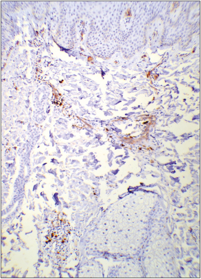
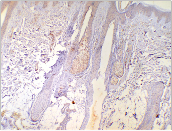
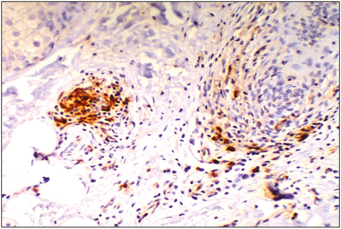
Classification of scarring alopecia poses a major problem, as there is considerable clinicopathologic overlap, particularly between lupus erythematosus (LE) and lichen planopilaris (LPP), especially in later stages. CD123 positive plasmacytoid dendritic cells (PDC) have been shown recently to be present in all forms of LE and are touted to be useful in differentiating LE from other scarring alopecias. Their distribution in non-scarring alopecia is not well documented. This is the first study that examines the PDC in both scarring and non-scarring alopecias.
Objective: To study the expression patterns of PDC in cases of both scarring and non-scarring alopecia.
Materials and methods: A total of 69 cases of alopecia (48 scarring, 21 non-scarring) were studied for CD123 expression by immunohistochemistry.
Results: Among the scarring alopecias, 17/20 LE cases showed PDC in contrast to 1/22 LPP cases. This difference was statistically significant (P = 0.0001). 1/2 cases of folliculitis decalvans showed PDC. None of the cases of unclassified scarring alopecia were positive. In the non-scarring group, 19/20 cases of alopecia areata and a single case of trichotillomania lacked PDC.
Conclusion: The finding of CD123 expressing PDC appears to be a promising parameter in distinguishing LE from other forms of alopecia.
Keywords: CD 123; lupus erythematosus; plasmacytoid dendritic cells; scarring alopecia.
Copyright: © 2022 Journal of Cutaneous and Aesthetic Surgery.
Conflict of interest statement
There are no conflicts of interest.
Figures
Similar articles
-
Value of CD123 Immunohistochemistry and Elastic Staining in Differentiating Discoid Lupus Erythematosus from Lichen Planopilaris.Int J Trichology. 2020 Mar-Apr;12(2):62-67. doi: 10.4103/ijt.ijt_32_20. Epub 2020 May 5. Int J Trichology. 2020. PMID: 32684677 Free PMC article.
-
Evaluation of the Diagnostic Value of Plasmacytoid Dendritic Cells in Differentiating the Lymphocytic Cicatricial Alopecias.Dermatology. 2015;231(2):158-63. doi: 10.1159/000431174. Epub 2015 Jun 17. Dermatology. 2015. PMID: 26088789
-
CD123 immunohistochemistry for plasmacytoid dendritic cells is useful in the diagnosis of scarring alopecia.J Cutan Pathol. 2016 Aug;43(8):643-8. doi: 10.1111/cup.12725. Epub 2016 Jun 3. J Cutan Pathol. 2016. PMID: 27130548
-
Histopathology of alopecia: a clinicopathological approach to diagnosis.Histopathology. 2010 Jan;56(1):24-38. doi: 10.1111/j.1365-2559.2009.03439.x. Histopathology. 2010. PMID: 20055903 Review.
-
Histologic features of alopecias: part II: scarring alopecias.Actas Dermosifiliogr. 2015 May;106(4):260-70. doi: 10.1016/j.ad.2014.06.016. Epub 2014 Oct 24. Actas Dermosifiliogr. 2015. PMID: 25439143 Review. English, Spanish.
Cited by
-
The role of plasmacytoid dendritic cells (pDCs) in immunity during viral infections and beyond.Cell Mol Immunol. 2024 Sep;21(9):1008-1035. doi: 10.1038/s41423-024-01167-5. Epub 2024 May 22. Cell Mol Immunol. 2024. PMID: 38777879 Free PMC article. Review.
-
Alopecias: Practical Tips for the Management of Biopsies and Main Diagnostic Clues for General Pathologists and Dermatopathologists.J Clin Med. 2023 Jul 29;12(15):5004. doi: 10.3390/jcm12155004. J Clin Med. 2023. PMID: 37568407 Free PMC article. Review.
References
-
- Tomasini D, Mentzel T, Hantschke M, Cerri A, Paredes B, Rütten A, et al. Plasmacytoid dendritic cells: An overview of their presence and distribution in different inflammatory skin diseases, with special emphasis on jessner’s lymphocytic infiltrate of the skin and cutaneous lupus erythematosus. J Cutan Pathol. 2010;37:1132–9. - PubMed
-
- McNiff JM, Kaplan DH. Plasmacytoid dendritic cells are present in cutaneous dermatomyositis lesions in a pattern distinct from lupus erythematosus. J Cutan Pathol. 2008;35:452–6. - PubMed
LinkOut - more resources
Full Text Sources