

Clinical and Dermoscopic Evaluation of Periorbital Melanosis
- PMID: 35965914
- PMCID: PMC9364450
- DOI: 10.4103/JCAS.JCAS_109_21
Clinical and Dermoscopic Evaluation of Periorbital Melanosis
Abstract
Background: Periorbital melanosis (POM) describes the light-to-dark-colored, brownish-black pigmentation surrounding the eyelids. It can affect an individual's quality of life. Dermoscopic features of POM are not frequently reported in the literature.
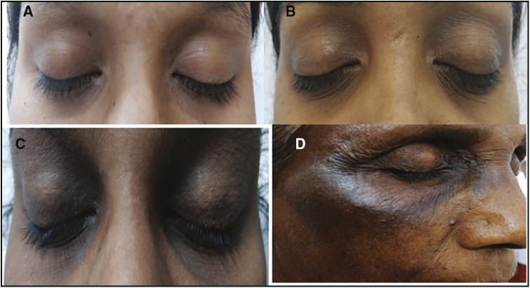
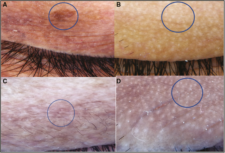
Materials and methods: This study comprised 100 patients aged above 16 years, who attended our outpatient department (OPD) from November 2018 to October 2019. A detailed history, clinical features, and the dermoscopic study of color, pattern of pigment, and pattern of the blood vessel were recorded with the Dermlite-3N dermoscope (3Gen, San Juan Capistrano, California). On the basis of the eyelids' pigmentation and involvement, patients were clinically graded as Grade 0 to 4, with 4 being deep dark color extending beyond the infraorbital fold. The clinical patterns and the dermoscopic features were correlated.
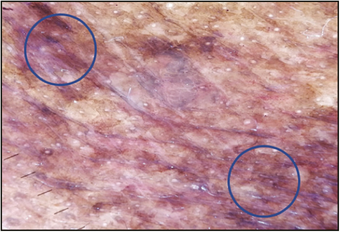
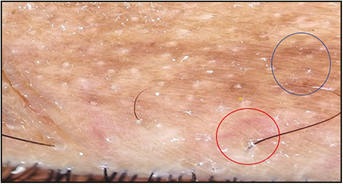
Results: Most patients were women (76) and the common age group was 16-25 years. Most of the patients had both the eyelids involved (58%), followed by lower eyelids (28%). The majority of the patients were having POM of grade 2 (47%). Seventeen patients (17%) had a positive family history of POM. The most common clinical form of POM observed was constitutional type (77) followed by postinflammatory type (12). Of 100 patients, 52 had pigmentary, 15 had vascular, and 33 had mixed pigmentary-vascular pattern. Cell phone usage (>4 h) and refractory errors (38% each) were the common risk factors observed. Stress and respiratory allergy were significantly associated. In the pigmentation patterns, epidermal (54%), dermal (14%), and mixed (17%) subsets were observed. The reticular pattern was the most common vascular pattern (65%).
Conclusion: POM is a multifactorial entity. Multiple risk factors play a role in the pathogenesis and aggravation. Clinical forms did not show any specific dermoscopic patterns. Dermoscopy of POM helps to know the underlying pathology, which in turn paves the way to the effective treatment.
Keywords: Dermoscopy; periorbital melanosis; pigmentary patterns; vascular patterns.
Copyright: © 2022 Journal of Cutaneous and Aesthetic Surgery.
Conflict of interest statement
There are no conflicts of interest.
Figures
Similar articles
-
A cross-sectional study on clinico-dermoscopic features of periorbital melanosis in a tertiary care hospital.J Cosmet Dermatol. 2021 Sep;20(9):2917-2923. doi: 10.1111/jocd.13979. Epub 2021 Feb 15. J Cosmet Dermatol. 2021. PMID: 33544960
-
Dermoscopy as an efficient aid to diagnose pigmentary and vascular component of periorbital melanosis: A cross-sectional study.J Cosmet Dermatol. 2022 Nov;21(11):5880-5886. doi: 10.1111/jocd.15141. Epub 2022 Jun 21. J Cosmet Dermatol. 2022. PMID: 35665587
-
Dermatoscopy of primary localised cutaneous amyloidosis - A cross-sectional study in a setting of South Asian public dermatology department.Skin Health Dis. 2023 Nov 27;4(1):e316. doi: 10.1002/ski2.316. eCollection 2024 Feb. Skin Health Dis. 2023. PMID: 38312259 Free PMC article.
-
Dermoscopy.J Dermatol. 2006 Aug;33(8):513-7. doi: 10.1111/j.1346-8138.2006.00126.x. J Dermatol. 2006. PMID: 16923131 Review.
-
Dermoscopy of acquired pigmentary disorders: a comprehensive review.Int J Dermatol. 2022 Jan;61(1):7-19. doi: 10.1111/ijd.15741. Epub 2021 Jul 7. Int J Dermatol. 2022. PMID: 34235719 Review.
References
-
- Ahuja SK, Deshmukh AR, Khushalani SR. A study of dermatoscopic pattern of periorbital hypermelanosis. PigmentInt. 2017;4:29–34.
-
- Jage M, Mahajan S. Clinical and dermoscopic evaluation of periorbital hyperpigmentation. Indian J Dermatopathol Diagn Dermatol. 2018;5:42–7.
-
- Strachan T, Read AP. Genes in pedigrees and population. In: Strachan T, editor. Human molecular genetics. 3rd ed. New York: Garland Science; 2003. pp. 106–7.
LinkOut - more resources
Full Text Sources