

Coronary artery intramural hematoma, a rare complication of percutaneous coronary intervention
- PMID: 35965935
- PMCID: PMC9364049
- DOI: 10.1016/j.radcr.2022.07.001
Coronary artery intramural hematoma, a rare complication of percutaneous coronary intervention
Abstract
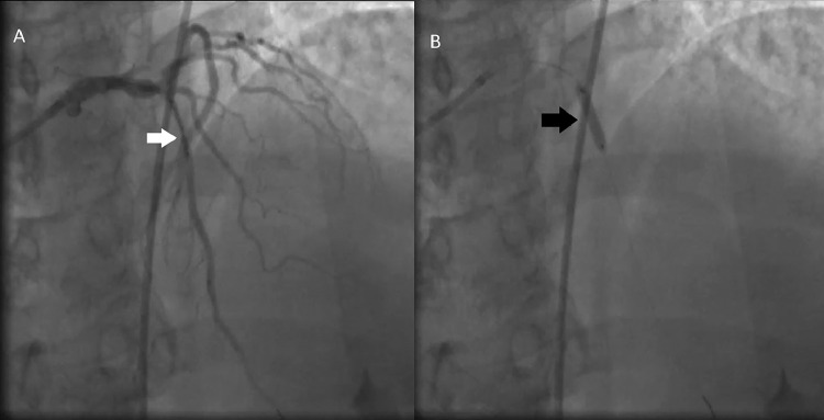
Coronary artery intramural hematoma is a rare complication of percutaneous coronary intervention which develops from intimal tear of coronary artery and propagates by blood accumulation along the medial surface of adjacent segment. Fifty-three-year-old male presented with nonexertional chest pain; he was referred after a positive stress test with+ moderate lateral wall ischemia. Coronary angiography showed 80% lesion in mid-left anterior descending artery (mLAD). Angiogram after angioplasty with 2.0 mm × 15 mm balloon and 3.0 mm × 15 mm drug-eluting-stent demonstrated a new stenotic lesion distal to stented mLAD segment. Subsequently, an overlapping 3.0 mm × 30 mm stent was placed with effective restoration of blood flow through LAD. During percutaneous coronary intervention (PCI), balloon predilatation can result in plaque fracture and stent deployment may cause intimal tear forming intramural hematoma which can lead to post-PCI myocardial infarction necessitating prompt detection by intravascular imaging with intravascular ultrasound and optical coherence tomography. Management is based on individual patient's characteristics and includes medical therapy, angiographic surveillance or repeat PCI.
Keywords: Angiogram; CIH, Coronary Intramural Hematoma; Coronary angiography; Coronary artery intramural hematoma; DES, Drug-eluting stent; ICU, Intensive care unit; IVUS, Intravascular Ultrasound; Iatrogenic myocardial infarction; LAD, left anterior descending artery; LVEF, left ventricular ejection fraction; OCT, Optical Coherence Tomography; PCI, Percutaneous Coronary Intervention; Percutaneous coronary intervention; Post-percutaneous coronary intervention myocardial infarction; mLAD, mid segment of left anterior descending artery.
© 2022 The Authors. Published by Elsevier Inc. on behalf of University of Washington.
Figures


Similar articles
-
Coronary Artery Occlusion Caused by Intramural Hematoma Due to In-Stent Dissection.JACC Case Rep. 2020 May 20;2(5):707-708. doi: 10.1016/j.jaccas.2020.03.011. eCollection 2020 May. JACC Case Rep. 2020. PMID: 34317330 Free PMC article.
-
Balloon rupture during coronary angioplasty causing dissection and intramural hematoma of the coronary artery; a case report.J Cardiol Cases. 2009 Nov 8;1(1):e17-e20. doi: 10.1016/j.jccase.2009.06.002. eCollection 2010 Feb. J Cardiol Cases. 2009. PMID: 30615766 Free PMC article.
-
An Uncommon Cause of ST-Segment Elevation Myocardial Infarction: Intramural Coronary Artery Hematoma After Blunt Chest Trauma.JACC Case Rep. 2020 Dec 16;2(15):2304-2309. doi: 10.1016/j.jaccas.2020.09.032. eCollection 2020 Dec. JACC Case Rep. 2020. PMID: 34317160 Free PMC article.
-
The Role of Vascular Imaging in Guiding Routine Percutaneous Coronary Interventions: A Meta-Analysis of Bare Metal Stent and Drug-Eluting Stent Trials.Cardiovasc Ther. 2015 Dec;33(6):360-6. doi: 10.1111/1755-5922.12160. Cardiovasc Ther. 2015. PMID: 26363283 Review.
-
Treatment strategies in the left main coronary artery disease associated with acute coronary syndromes.J Saudi Heart Assoc. 2015 Oct;27(4):272-6. doi: 10.1016/j.jsha.2015.03.002. Epub 2015 Mar 21. J Saudi Heart Assoc. 2015. PMID: 26557745 Free PMC article. Review.
Cited by
-
Management of Catheter-Induced Coronary Artery Dissection Leading to Extensive Bidirectional Intramural Hematoma.JACC Case Rep. 2025 Mar 5;30(5):103173. doi: 10.1016/j.jaccas.2024.103173. JACC Case Rep. 2025. PMID: 40054951 Free PMC article.
References
-
- Maehara A, Mintz GS, Bui AB, et al. Incidence, morphology, angiographic findings, and outcomes of intramural hematomas after percutaneous coronary interventions. Circulation. 2002;105:2037–2042. doi: 10.1161/01.CIR.0000015503.04751.BD. - DOI - PubMed
-
- Mintz GS, Nissen SE, Anderson WD, et al. American College of Cardiology Clinical Expert Consensus document on standards for acquisition, measurement and reporting of Intravascular Ultrasound Studies (IVUS). A report of the American College of Cardiology Task Force on Clinical Expert Consensus Documents. J Am Coll Cardiol. 2001;37:1478–1492. doi: 10.1016/S0735-1097(01)01175-5. - DOI - PubMed
Publication types
LinkOut - more resources
Full Text Sources
Miscellaneous
