

Case report: Utilization of neutral density filters for densitometry analysis of dense corneal opacities
- PMID: 35966118
- PMCID: PMC9364089
- DOI: 10.1016/j.ajoc.2022.101672
Case report: Utilization of neutral density filters for densitometry analysis of dense corneal opacities
Abstract
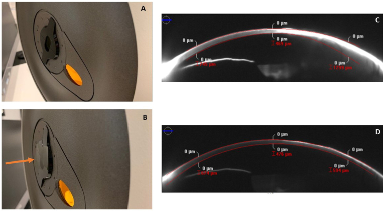
Purpose: This report describes the technique of utilizing a neutral density filter (NDF) during Scheimpflug imaging of a dense corneal opacity in order to increase data acquisition success and improve data reliability for densitometry analysis.
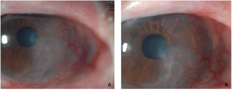
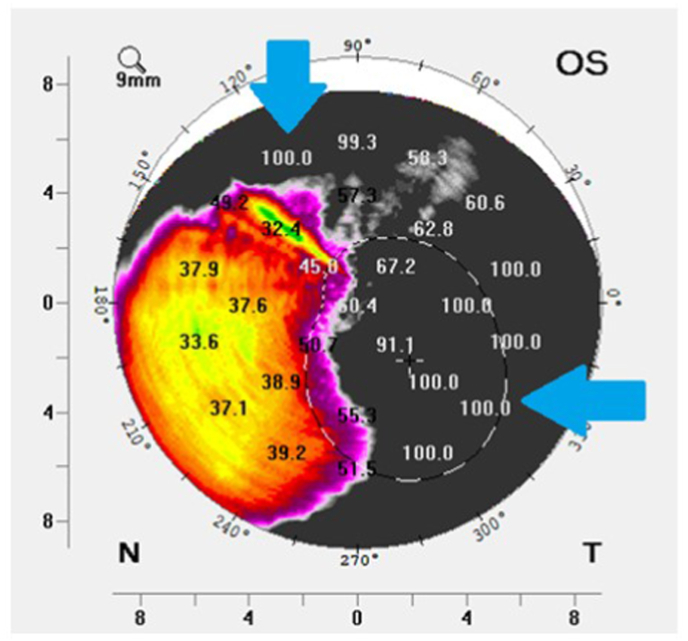
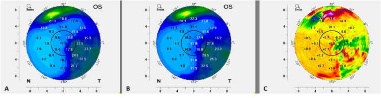
Observations: A 49-year-old female with Steven-Johnson Syndrome secondary to sulfonamide use presented for routine follow up evaluation of her customized ocular surface prosthetic device (PD). Her ocular history was significant for mucous membrane grafting and limbal stem cell transplant in both eyes. The ocular surface examination of the left eye was notable for chronic dense neovascularization and scarring of the temporal and inferior cornea which extended into the visual axis. Scheimpflug imaging and densitometry analysis were performed in order to quantify the severity of the scar, however, there was significant difficulty in acquiring densitometry data. During a subsequent follow-up visit to monitor the scar, standardized room lighting and a neutral density filter were used to obtain reproducible and reliable imaging for densitometry analysis. The corneal scar was monitored over time using this standardized imaging protocol and by densitometry analysis minimal progression of the scar was evident, suggesting that recently documented significant vision loss in the left eye could not be attributed solely to changes in the scar.
Conclusion and importance: The use of a neutral density filter along with standardized ambient lighting conditions when performing Scheimpflug imaging may be necessary to reliably monitor densitometry progression of clinically severe corneal opacities.
Keywords: Corneal scar; Scheimpflug imaging; Scleral lens.
© 2022 The Authors.
Conflict of interest statement
The authors report no financial conflicts of interest in this work. Daniel Brocks is a salaried clinical employee of BostonSight, Needham, MA. None of the authors have a propriety or financial interest in PROSE (BostonSight, Needham MA) or the prosthetic devices used in PROSE treatment.
Figures
Similar articles
-
Limbal stem cell transplantation: an evidence-based analysis.Ont Health Technol Assess Ser. 2008;8(7):1-58. Epub 2008 Oct 1. Ont Health Technol Assess Ser. 2008. PMID: 23074512 Free PMC article.
-
A Case of Artificial Snow Foam induced Corneal Endotheliitis Followed up by Scheimpflug Densitometry.Med Hypothesis Discov Innov Ophthalmol. 2019 Summer;8(2):64-68. Med Hypothesis Discov Innov Ophthalmol. 2019. PMID: 31263714 Free PMC article.
-
How Does Light Intensity of the Recording Room Affect the Evaluation of Lens and Corneal Clarity by Scheimpflug Tomography?Cornea. 2020 Feb;39(2):137-139. doi: 10.1097/ICO.0000000000002212. Cornea. 2020. PMID: 31714403
-
Corneal structure, transparency, thickness and optical density (densitometry), especially as relevant to contact lens wear-a review.Cont Lens Anterior Eye. 2019 Jun;42(3):238-245. doi: 10.1016/j.clae.2018.11.014. Epub 2018 Nov 28. Cont Lens Anterior Eye. 2019. PMID: 30502960 Review.
-
[Image analysis and Sheimpflug photography of anterior segment of the eye--a review].Klin Monbl Augenheilkd. 2001 Feb;218(2):67-77. doi: 10.1055/s-2001-12248. Klin Monbl Augenheilkd. 2001. PMID: 11258128 Review. German.
References
-
- Parsons J.H. Parson's Diseases of the Eye. Elsevier, A Division of Reed Elsevier India Private Limited; New Delhi: 2011. Diseases of the cornea; p. 190.
-
- Shimizu E., Yamaguchi T., Tsubota K., Shimazaki J. Corneal higher-order aberrations in eyes with corneal scar after traumatic perforation. Eye Contact Lens. 2019;45(2):124–131. - PubMed
Publication types
LinkOut - more resources
Full Text Sources
